
Building upon over twenty years of investigation, a research project by MIT neuroscientists at The Picower Institute for Learning and Memory presents a novel method to address the pathology and manifestations of fragile X syndrome, which is the most prevalent genetically-induced autism spectrum disorder. The group demonstrated that enhancing a new form of neurotransmitter signaling diminished key characteristics of fragile X in murine models of the disorder.
The innovative technique, outlined in Cell Reports, operates by focusing on a particular molecular subunit of “NMDA” receptors which they found to be crucial in how neurons produce proteins to manage their connections, or “synapses,” with other neurons in neural circuits. The researchers illustrated that in fragile X model mice, increasing the receptor’s activity prompted neurons in the hippocampus area of the brain to enhance molecular signaling that inhibited excessive bulk protein production, resulting in other significant improvements.
Establishing the foundation
“One aspect I find particularly rewarding about this research is how well the pieces of the puzzle integrate with earlier findings,” remarks study senior author Mark Bear, Picower Professor in MIT’s Department of Brain and Cognitive Sciences. Former postdoctoral researcher Stephanie Barnes, now an instructor at the University of Glasgow, serves as the primary author of the study.
Bear’s laboratory investigates how neurons constantly modify their circuit connections, a mechanism known as “synaptic plasticity,” which scientists believe underpins the brain’s capability to adapt to experiences and to form and recall memories. These investigations led to two findings that laid the groundwork for the recent published breakthroughs. In 2011, Bear’s lab demonstrated that fragile X and another autism disorder, tuberous sclerosis (Tsc), represented opposite ends of a spectrum concerning a specific kind of protein synthesis occurring in the same neurons. In fragile X, there was an excess, while in Tsc, there was a deficiency. When lab members interbred fragile X and Tsc mice, their offspring turned out to be healthy, as the mutations from each disorder effectively negated each other.
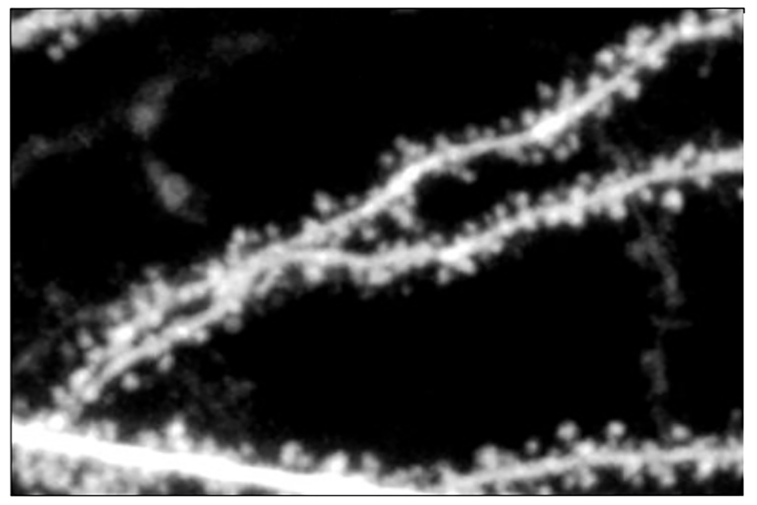
More recently, Bear’s team revealed a different dichotomy. Their influential work from the 1990s established that the passage of calcium ions through NMDA receptors can instigate a form of synaptic plasticity known as “long-term depression” (LTD). However, in 2020, they discovered that an alternative mode of signaling by the receptor — one which did not necessitate ion flow — modified protein synthesis in the neuron and resulted in a physical reduction of the dendritic “spine” structures that accommodate synapses.
For Bear and Barnes, these findings suggested that if they could determine how NMDA receptors influence protein synthesis, they might uncover a new mechanism that could be therapeutically adjusted to tackle fragile X (and possibly tuberous sclerosis) pathology and symptoms. This would represent a significant advancement complementing ongoing research undertaken by Bear’s lab to rectify fragile X protein synthesis levels via another receptor known as mGluR5.
Dissection of the receptor
In the current study, Bear and Barnes’ team opted to utilize the non-ionic effect on spine shrinkage as an indicator to dissect how NMDARs influence protein synthesis for synaptic plasticity in hippocampal neurons. They theorized that the discrepancy between ionic effects on synaptic functionality and non-ionic effects on spine structure might originate from the existence of two separate components of NMDA receptors: “subunits” identified as GluN2A and GluN2B. To validate this, they employed genetic alterations to eliminate each of the subunits. Upon doing so, they discovered that the removal of either “2A” or “2B” could abolish LTD, though only the removal of 2B influenced spine dimensions. Further investigations revealed that both 2A and 2B are necessary for LTD, but the reduction in spine size is solely reliant on the 2B subunit.
The subsequent challenge was to clarify how the 2B subunit signals spine contraction. A promising suspect was a portion of the subunit known as the “carboxyterminal domain,” or CTD. Therefore, in a new experiment, Bear and Barnes utilized a mouse genetically modified by researchers at the University of Edinburgh, allowing for the swap of the 2A and 2B CTDs. A revealing outcome was observed when the 2B subunit lacked its appropriate CTD, resulting in the elimination of its effect on spine structure. This finding confirmed that the 2B subunit signals spine reduction through its CTD.
Another outcome of replacing the CTD within the 2B subunit was an increase in bulk protein synthesis resembling findings in fragile X. Conversely, enhancing the non-ionic signaling through the 2B subunit decreased bulk protein synthesis, akin to Tsc.
Addressing fragile X
Piecing the findings together indicated that augmenting signaling through the 2B subunit may, similar to introducing the mutation causing Tsc, restore certain aspects of fragile X.
In fact, when the researchers incorporated the 2B subunit CTD of the NMDA receptor in fragile X model mice, they observed restoration not only of excessive bulk protein synthesis but also adjustments in synaptic plasticity and increased electrical excitability that characterize the disease. To gauge the potential effectiveness of a treatment focusing on NMDA receptors for fragile X, they experimented with an investigational drug known as Glyx-13. This pharmaceutical binds to the 2B subunit of NMDA receptors to enhance signaling. The team discovered that this treatment could also normalize protein synthesis and diminish sound-induced seizures in fragile X mice.
Based on another preceding study conducted in the lab, the researchers now hypothesize that the favorable impact of the 2B subunit’s CTD signaling in fragile X mice is that it modifies the protein synthesis balance away from an overly efficient translation of short messenger RNAs (which causes excessive bulk protein production) toward a less efficient translation of longer messenger RNAs.
Bear states he is uncertain about the prospects for Glyx-13 as a clinical treatment, but he acknowledged that there are several drugs in clinical development explicitly targeting the 2B subunit of NMDA receptors.
Alongside Bear and Barnes, the study’s additional authors include Aurore Thomazeau, Peter Finnie, Max Heinreich, Arnold Heynen, Noboru Komiyama, Seth Grant, Frank Menniti, and Emily Osterweil.
The FRAXA Foundation, The Picower Institute for Learning and Memory, The Freedom Together Foundation, and the National Institutes of Health financed this research.