
“`html
Science & Tech
Unraveling the evolutionary enigma of how humans began to walk upright
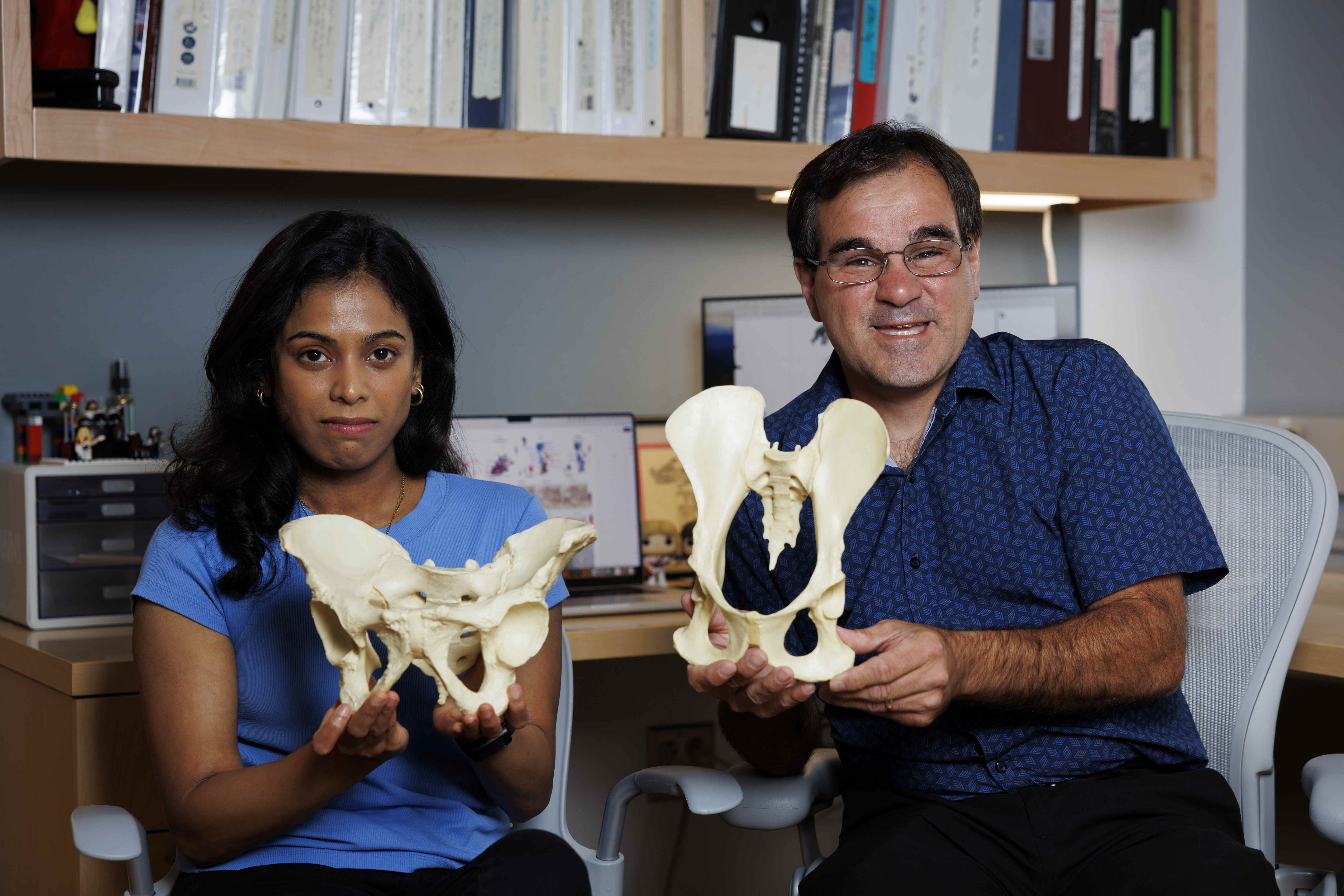
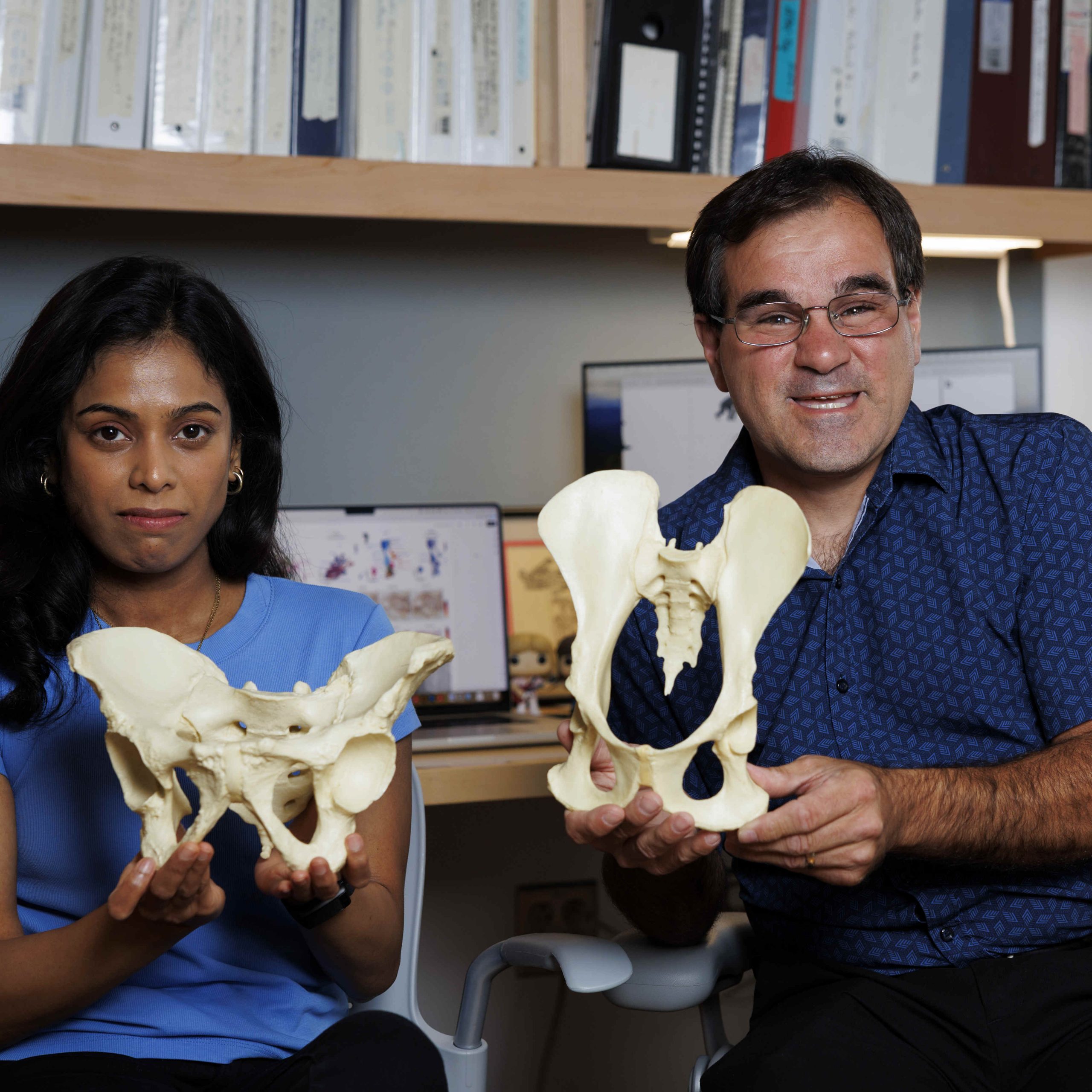
Gayani Senevirathne (left) displays the shorter, broader human pelvis, which evolved from the longer upper hipbones of primates, presented by Terence Capellini.
Niles Singer/Harvard Staff Photographer
New research uncovers genetic and developmental transformations that reshaped the pelvis, distinguishing ancestors from other primates
The pelvis is frequently referred to as the cornerstone of upright movement. Unlike any other aspect of our lower anatomy, it has undergone significant modifications throughout millions of years, enabling our forebears to evolve into the bipeds who explored and inhabited the Earth.
However, the mechanism by which evolution effectuated this drastic transformation has been elusive. A recent study published in the journal Nature, spearheaded by Harvard researchers, unveils two pivotal genetic alterations that reshaped the pelvis and facilitated our peculiar tendency to walk on two legs.
“Our work illustrates a complete mechanistic transformation in human evolution,” stated Terence Capellini, professor and chair of the Department of Human Evolutionary Biology and senior author of the study. “There is nothing comparable in other primates. The evolution of innovation — like the metamorphosis from fins to limbs or the formation of bat wings from fingers — typically involves significant changes in how developmental processes operate. In this case, we observe humanity undergoing a similar phenomenon, specifically concerning their pelves.”
Anatomists have long recognized that the human pelvis stands out among primates. The upper hipbones, or ilia, of chimpanzees, bonobos, and gorillas — our closest relatives — are tall, slender, and aligned flat from front to back. When viewed from the side, they resemble elongated blades. The configuration of the ape pelvis supports substantial muscles essential for climbing.
In contrast, the human hipbones have rotated sideways to create a bowl shape (in fact, the term “pelvis” is derived from the Latin word for basin). Our spreading hipbones allow for muscle attachments that help us maintain equilibrium as we shift our weight while walking and running.
In their latest publication, the collaborative group of researchers pinpointed some of the primary genetic and developmental transformations that thoroughly reformed the quadrupedal ape pelvis into one adapted for bipedality.
“Our goal was to synthesize various methodologies to construct a comprehensive narrative about the pelvic development over time,” noted Gayani Senevirathne, a postdoctoral fellow in Capellini’s lab and lead author of the study.
Senevirathne examined 128 samples of embryonic tissues from humans and nearly twenty other primate species housed in museums across the U.S. and Europe. These collections included specimens over a century old, preserved on glass slides or in jars.
The researchers also investigated human embryonic tissues obtained from the Birth Defects Research Laboratory at the University of Washington. They performed CT scans and scrutinized histology (the microscopic structure of tissues) to elucidate the pelvic anatomy during its early developmental phases.
“The work Gayani conducted was remarkable,” Capellini stated. “This was akin to five projects rolled into one.”
The researchers found that evolution restructured the human pelvis in two principal phases. Initially, a growth plate was rotated by 90 degrees to render the human ilium broad rather than tall. Subsequently, another shift modified the developmental timeline of bone formation.
Most lower body bones form through a mechanism that commences when cartilage cells develop on growth plates aligned along the length of the growing bone. This cartilage subsequently hardens into bone through a process known as ossification.
In the early phases of development, the human iliac growth plate formed with growth aligned head-to-tail, similar to other primates. However, by day 53, the growth plates in humans evolved to dramatically shift perpendicularly from the original alignment — thereby shortening and widening the hipbone.
“When observing the pelvis, that hadn’t crossed my mind,” Capellini commented. “I expected a gradual progression of shortening first and then widening. However, the histology truly indicated that it actually rotated 90 degrees — causing it to become short and wide concurrently.”
The authors propose that these modifications commenced with the reorientation of growth plates around the time our ancestors diverged from the African apes, estimated to have occurred between 5 million and 8 million years ago.
Another significant alteration pertained to the timing of bone formation.
Most bones develop from a primary ossification center situated in the center of the bone shaft.
In humans, however, the ilia exhibit a distinctive pattern. Ossification initiates at the back of the sacrum and radiates outward. This mineralization is confined to the outer layer, with interior ossification lagging by 16 weeks in comparison to other primates — allowing the bone to retain its shape during growth and fundamentally altering its geometry.
“Embryonically, by 10 weeks, you already have a pelvis,” said Capellini while sketching on a whiteboard. “It appears like this — basin-shaped.”
To uncover the molecular mechanisms driving this transformation, Senevirathne utilized techniques such as single-cell multiomics and spatial transcriptomics. The team identified over 300 active genes, including three with significant influence — SOX9 and PTH1R (regulating the growth plate rotation) and RUNX2 (governing the change in ossification).
The significance of these genes became evident in disorders caused by their dysfunction. For instance, a mutation in SOX9 leads to campomelic dysplasia, a condition resulting in hipbones that are abnormally narrow and lack lateral flaring.
Likewise, mutations in PTH1R result in excessively narrow hipbones and other skeletal disorders.
The authors speculate that these transformations began with the reorientation of growth plates around the time our ancestors diverged from the African apes, estimated to have transpired between 5 million and 8 million years ago.
They surmise that the pelvis remained a crucial site of evolutionary change for millions of years.
As cranial sizes increased, the pelvis experienced another selective pressure termed the “obstetrical dilemma” — the compromise between a narrow pelvis (beneficial for efficient movement) and a wider one (facilitating the delivery of larger-brained offspring).
They propose that the delayed ossification likely emerged in the last 2 million years.
The most ancient pelvis discovered in the fossil record is the 4.4-million-year-old Ardipithecus from Ethiopia (a hybrid of an upright walker and tree climber with a grasping toe), displaying early signs of human-like characteristics in the pelvis.
The renowned 3.2-million-year-old Lucy skeleton, also from Ethiopia, encompasses a pelvis that exhibits further evolution of bipedal traits like flaring hip blades suitable for bipedal musculature.
Capellini believes the new findings should inspire scientists to reevaluate some fundamental beliefs regarding human evolution.
“All fossil hominids from that era were developing the pelvis in a manner unlike any other primate that preceded them,” remarked Capellini. “Later increases in brain size should not be assessed through a growth model akin to that of chimpanzees and other primates. The model should be based on the evolutions occurring in humans and hominins. The subsequent growth of fetal head size transpired against the backdrop of a newly established manner of forming the pelvis.”
This study was partially financed by the National Institutes of Health.
“`