
Whether you’re a social butterfly or a secluded soul, life has a way of presenting various situations. What you require is a nervous system adaptable enough to navigate these challenges. In a recent investigation, MIT neuroscientists revealed how even a basic organism can reorganize neural circuits and the chemical messengers, or “neuromodulators,” in its brain to mount a flexible response to an infection. This study may thus offer insight into how brains in more intricate organisms, including humans, utilize their available resources to adjust to fluctuating internal conditions.
“Neuromodulators serve crucial functions in linking alterations in animals’ internal conditions to their actions,” the researchers assert in their manuscript, which was recently published in Nature Communications. “How different combinations of neuromodulators released from various neuronal origins regulate the wide range of internal conditions exhibited by animals remains an unresolved issue.”
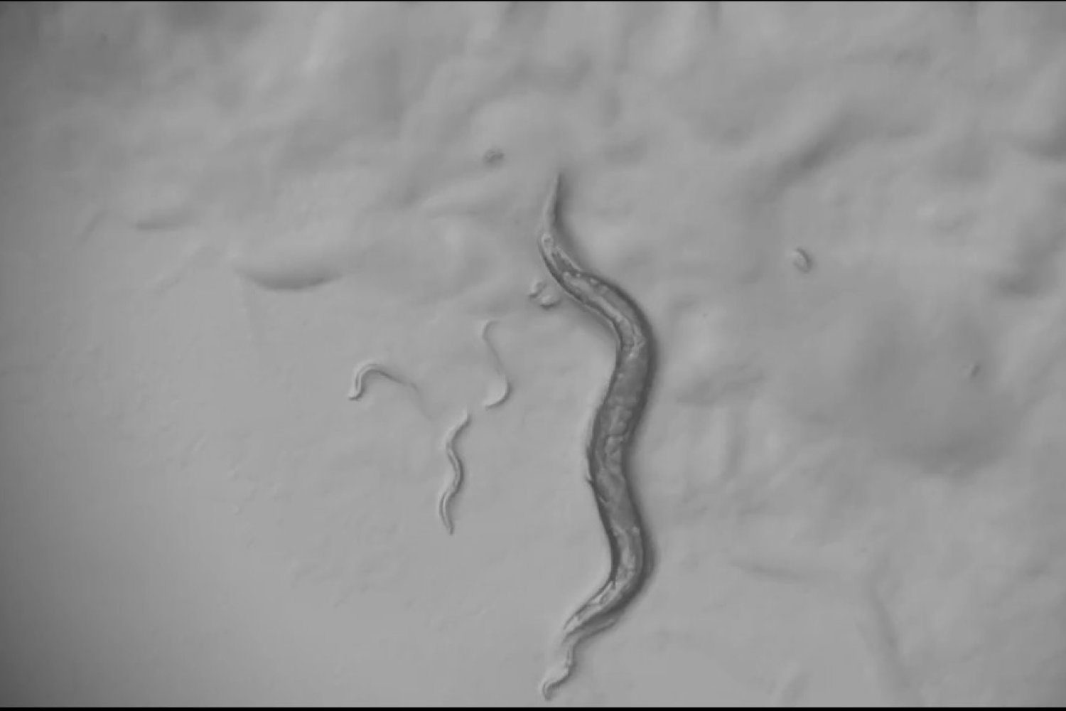
When C. elegans worms consumed infectious Pseudomonas bacteria, their food intake diminished and they became increasingly sluggish. Upon examining the nervous system to understand this behavior, the scientists identified that the worm had entirely transformed the functions of numerous neurons within its 302 neurons and some of the peptides they secrete throughout the brain to influence behavior. Systems that previously responded to stress in one scenario and fullness in another were restructured to tackle the infection.
“The essence of the inquiry is about how you acclimate to your surroundings with the greatest degree of flexibility using the set of neurons and neuromodulators at your disposal,” states postdoc Sreeparna Pradhan, co-lead author of the new study in Nature Communications. “How do you maximize the range of options available?”
The investigation was conducted in the lab of senior author Steve Flavell, an associate professor in The Picower Institute for Learning and Memory and the Department of Brain and Cognitive Sciences, as well as an investigator at the Howard Hughes Medical Institute. Pradhan, who received a fellowship from MIT’s K. Lisa Yang Brain-Body Center during this work, collaborated with former Flavell Lab graduate student Gurrein Madan to spearhead the research.
Pradhan mentions that the team encountered several unexpected findings throughout the study, including that a neuropeptide named FLP-13 completely altered its role in infected worms compared to those under different stress conditions. Earlier research indicated that when worms experience heat stress, a neuron known as ALA releases FLP-13 to induce a quiescent state. Yet, when the worms in this study ingested Pseudomonas bacteria, a network of different neurons discharged FLP-13 to counter quiescence, allowing the worms to endure longer. Concurrently, ALA assumed a different role during illness: spearheading the effort to curb feeding by releasing a distinct group of peptides.
A holistic approach
To comprehend how the worms reacted to infection, the team monitored an array of behavioral traits for days and performed genetic modifications to explore the fundamental mechanisms involved. They also captured activity across the worms’ entire brains. This level of comprehensive observation and experimentation is challenging in more complex organisms, but Pradhan suggests that the relative simplicity of C. elegans makes it a practical model organism. The team’s strategy also enabled them to uncover many unexpected results.
For example, Pradhan didn’t initially anticipate that the ALA neuron would be the one to suppress feeding, but after observing their behavior over time, she began to notice that the reduced intake stemmed from the worms taking brief pauses they typically wouldn’t. As she and Madan manipulated more than a dozen genes they believed could impact behavior and feeding in the worms, she included another gene called ceh-17 that she had learned about years prior, which appeared to encourage episodes of “microsleep” in the worms. Upon knocking out ceh-17, they observed that those worms did not reduce feeding when infected, unlike the typical subjects. It turned out that ceh-17 is crucial for ALA’s proper function, leading the team to conclude that ALA might play a role in the feeding-reduction behavior.
To confirm this, they subsequently knocked out various peptides released by ALA and found that when they eliminated three specific ones — flp-24, nlp-8, and flp-7 — the infected worms did not exhibit decreased feeding upon infection. This confirmed that ALA is responsible for driving the reduced feeding behavior by releasing those three peptides.
Additionally, Pradhan and Madan’s evaluations indicated that when infected worms lacked flp-13, they entered a quiescent state much sooner than infected worms that had the peptide. Notably, the worms that resisted the quiescent state had a longer lifespan. They found that this resistance depended on FLP-13 being produced by four neurons (I5, I1, ASH, and OLL), but not from ALA. Further tests revealed that FLP-13 interacted with a widespread neuropeptide receptor known as DMSR-1 to inhibit quiescence.
Taking a brief rest
The final significant revelation of the study was that the quiescent state induced by Pseudomonas infection in worms is distinct from other types of sleepiness triggered by different contexts, such as after fullness or heat stress. In those situations, the worms do not awaken easily (with a slight poke), but during infection, their quiescent state was easily reversible. It appeared more akin to lethargy than true sleep. Utilizing the lab’s capability to monitor all neural activity during various behaviors, Pradhan and Madan determined that a neuron named ASI was notably active during these episodes of lethargy. This observation was further solidified when they demonstrated that ASI’s secretion of the peptide DAF-7 was necessary for the quiescent state to manifest in infected animals.
In summary, the study illustrated that the worms adaptively repurpose and reconfigure — sometimes even completely reversing — the functions of neurons and peptides to respond to infections, in contrast to alternative challenges like stress. The findings therefore illuminate what has been a perplexing question. How do brains leverage their collection of cells, circuits, and neuromodulators to respond to life’s demands? A portion of the answer seems to involve reshuffling existing components rather than inventing new ones for each circumstance.
“The conditions of stress, satiety, and infection are not driven by distinct sets of neuromodulators,” the authors noted in their paper. “Instead, a broader array of neuromodulators may be used from different origins and in various combinations to delineate these different internal states.”
In addition to Pradhan, Madan, and Flavell, the paper includes contributions from Di Kang, Eric Bueno, Adam Atanas, Talya Kramer, Ugur Dag, Jessica Lage, Matthew Gomes, Alicia Kun-Yang Lu, and Jungyeon Park.
Funding for the research was provided by the Picower Institute, the Freedom Together Foundation, the K. Lisa Yang Brain-Body Center, and the Yang Tan Collective at MIT; the National Institutes of Health; the McKnight Foundation; the Alfred P. Sloan Foundation; and the Howard Hughes Medical Institute.