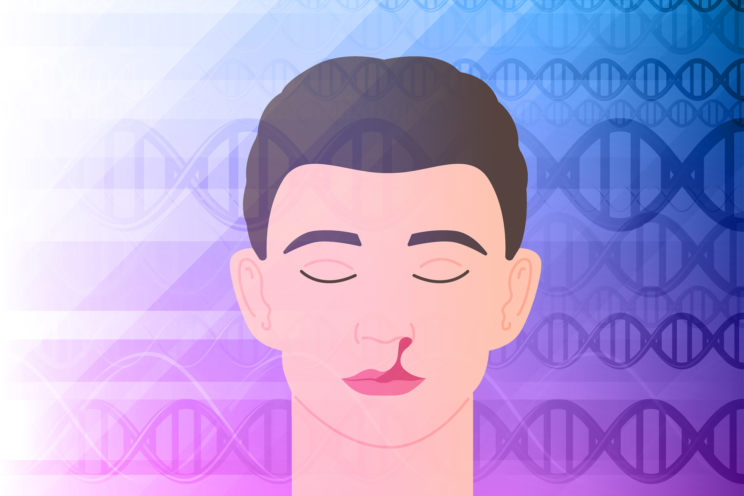
Cleft lip and cleft palate rank among the most prevalent congenital anomalies, appearing in approximately one in 1,050 births in the U.S. These anomalies arise when the tissues responsible for forming the lip or the mouth’s upper surface fail to unite completely, and they are thought to result from a combination of genetic and environmental influences.
In a recent investigation, biologists at MIT have uncovered how a genetic variant frequently observed in individuals with these facial deformities contributes to the onset of cleft lip and cleft palate.
Their research indicates that the variant reduces the availability of transfer RNA within cells, a crucial molecule required for protein assembly. Consequently, when this occurs, embryonic facial cells cannot merge to create the lip and the mouth’s roof.
“Up to now, no one had established the link that we did. This specific gene was known to be part of the intricate mechanism involved in the splicing of transfer RNA, but it wasn’t evident that it played such an essential role in this process and in facial development. In the absence of the gene, named DDX1, certain transfer RNA can no longer transport amino acids to the ribosome for new protein synthesis. If the cells are unable to properly manage these tRNAs, the ribosomes can no longer produce proteins,” states Michaela Bartusel, an MIT researcher and the primary author of the study.
Eliezer Calo, an associate professor of biology at MIT, serves as the senior author of the article, which is published today in the American Journal of Human Genetics.
Genetic Variants
Cleft lip and cleft palate, also referred to as orofacial clefts, can stem from genetic mutations, though in numerous instances, the genetic origins are unknown.
“The process underlying the formation of these orofacial clefts remains ambiguous, primarily because it is known that both genetic and environmental factors play a role,” Calo remarks. “Identifying what might be impacted has proven to be quite challenging in this scenario.”
To identify genetic elements influencing a specific disease, researchers typically conduct genome-wide association studies (GWAS), which can highlight variants that are observed more frequently in individuals afflicted with a particular illness compared to those who are not.
Concerning orofacial clefts, several genetic variants that consistently appear in GWAS seem to reside in a DNA segment that is non-coding. In this investigation, the MIT team aimed to elucidate how variants in this area might affect the emergence of facial deformities.
Their research indicated that these variants are situated in an enhancer region designated e2p24.2. Enhancers are sequences of DNA that interact with protein-coding genes, facilitating their activation by binding to transcription factors that promote gene expression.
The researchers discovered that this area is near three genes, implying that it could regulate the expression of those genes. One of these genes had already been excluded as a contributor to facial deformities, while another had previously shown a connection. In this investigation, the researchers concentrated on the third gene, known as DDX1.
It was found that DDX1 is essential for splicing transfer RNA (tRNA) molecules, which are vital for protein synthesis. Each tRNA molecule carries a specific amino acid to the ribosome — a cellular structure that assembles amino acids into proteins, directed by the information conveyed by messenger RNA.
Although around 400 distinct tRNAs can be found in the human genome, only a small subset of those requires splicing, and these are the tRNAs most impacted by the deficiency of DDX1. These tRNAs transport four different amino acids, and the researchers propose that these amino acids may be particularly important in proteins that embryonic cells need to develop the face effectively.
When the ribosomes require one of those four amino acids but none are accessible, the ribosome may halt, preventing protein synthesis.
The researchers are currently investigating which proteins could be most affected by the absence of those amino acids. They also intend to explore what occurs within cells when ribosomes stall, aiming to identify a stress signal that might be inhibited and assist cells in surviving.
Impaired tRNA
While this is the inaugural study connecting tRNA to craniofacial deformities, prior research has indicated that mutations disrupting ribosome assembly can also result in similar anomalies. Studies have additionally demonstrated that disruptions in tRNA synthesis — caused by mutations in the enzymes that attach amino acids to tRNA or in proteins involved in earlier stages of tRNA splicing — can lead to neurodevelopmental issues.
The researchers now aspire to investigate whether environmental influences connected to orofacial congenital anomalies also affect tRNA functionality. Preliminary findings suggest that oxidative stress — an accumulation of detrimental free radicals — may cause fragmentation of tRNA molecules. This oxidative stress can arise in embryonic cells following exposure to ethanol, as seen in fetal alcohol syndrome or if the mother develops gestational diabetes.
“I believe it is worthwhile to search for mutations that could contribute to this from a genetic perspective, and in the future, we will expand this investigation to consider which environmental factors influence tRNA functionality, and then determine which precautions might mitigate any negative effects on tRNAs,” states Bartusel.
The research received support from the National Science Foundation Graduate Research Program, the National Cancer Institute, the National Institute of General Medical Sciences, and the Pew Charitable Trusts.

