
Visualize a scenario where you wish to comprehend a movie’s storyline, but you’re limited to either visuals or audio. If you rely solely on visuals, you’ll overlook all the dialogues. Conversely, with audio alone, the action will be absent. Grasping our biology can be quite similar. Analyzing a certain type of data — for instance, which genes are active — can provide insights, but it represents merely one element of a complex narrative. To fully comprehend numerous biological processes and disease mechanisms, it is often necessary to merge various data types.
However, obtaining both the “visuals and audio” of biological data, such as gene activity and cell architecture from the same cells, compels researchers to formulate innovative strategies. It’s also essential for them to ensure that the gathered data accurately mirroring what transpires in living organisms, including the interactions between cells and their surroundings.
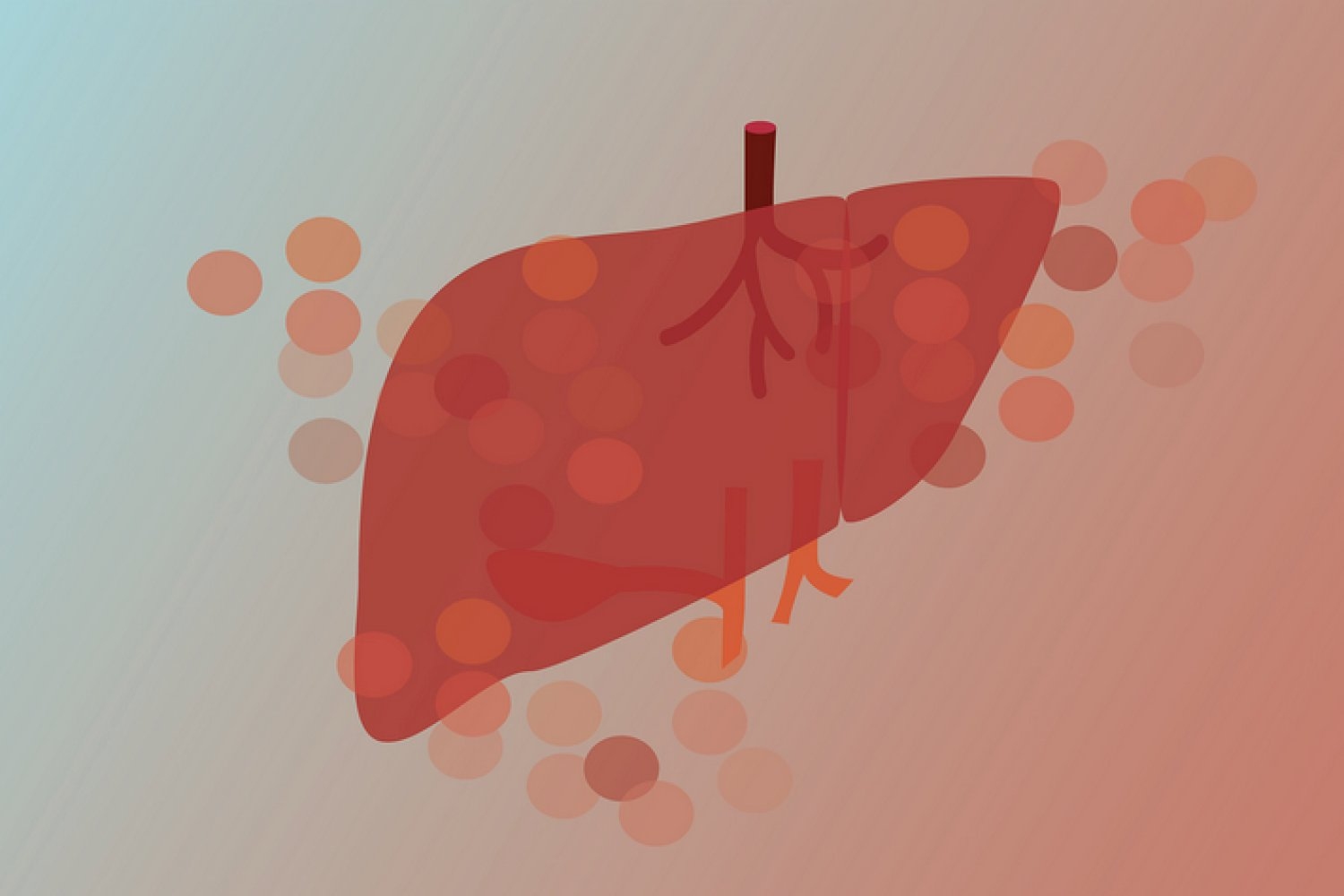
Researchers from the Whitehead Institute for Biomedical Research and Harvard University have tackled these challenges by developing Perturb-Multimodal (Perturb-Multi), a robust novel method that simultaneously gauges how genetic modifications, like silencing specific genes, influence both gene activity and cell architecture within intact liver tissue. This technique, detailed in Cell on June 12, aspires to hasten the understanding of how genes manage organ functionality and disease.
The research group, directed by Whitehead Institute Member Jonathan Weissman and his then-graduate student Reuben Saunders, along with Xiaowei Zhuang, the David B. Arnold Professor of Science at Harvard University, and her former postdoctoral researcher Will Allen, developed a system capable of testing numerous genetic modifications within a single mouse liver while acquiring multiple data types from the same cells.
“Grasping organ functionality necessitates examining various facets of cell biology simultaneously,” states Saunders. “With Perturb-Multi, we can observe how deactivating certain genes alters not only the activity of other genes but also the distribution of proteins within cells, the organization of cellular structures, and the positioning of cells in the tissue. It’s akin to having numerous specialized microscopes all concentrating on the same experiment.”
“This method expedites discovery by allowing us to assess the functions of numerous genes at once, and for each gene, it enables us to measure a variety of functional outputs or cellular properties concurrently — all within intact tissue from living specimens,” notes Zhuang, also a Howard Hughes Medical Institute (HHMI) investigator.
A more effective strategy for genetic investigations
Conventional genetic studies in mice frequently involve turning off a single gene and then observing the changes resulting from its absence to uncover the gene’s function. The researchers crafted their method to deactivate hundreds of genes throughout a single liver, ensuring that only one gene is turned off per cell — employing what’s termed a mosaic approach. This allowed them to examine the functions of numerous individual genes simultaneously in a single entity. They subsequently gathered a wide array of data from cells across the same liver to develop a comprehensive understanding of the repercussions of gene deactivation.
“Each cell acts as an independent experiment, and because all the cells originate from the same animal, we mitigate the variability introduced by comparing different mice,” remarks Saunders. “Every cell undergoes the same physiological conditions, nutrition, and environment, rendering our comparisons significantly more accurate.”
“The challenge we encountered is that tissues, to fulfill their roles, depend on thousands of genes expressed within various cells, collaborating in harmony. Each gene, in turn, can influence numerous aspects of a cell’s function. Testing these hundreds of genes in mice with existing techniques would be exceedingly slow and costly — practically impossible,” adds Allen.
Unveiling new biology through integrated measurements
The team utilized Perturb-Multi to investigate the genetic regulation of liver physiology and function. Their research yielded discoveries in three crucial areas of liver biology: hepatic fat accumulation — a precursor to liver disease; stress responses; and hepatocyte zonation (the specialization of liver cells, adopting different traits and functions based on their locations within the liver).
A notable finding emerged from examining genes that, when altered, induce fat accumulation in liver cells. Imaging data illustrated that four distinct genes resulted in similar fat droplet accumulation, while sequencing data revealed they did so via three entirely different mechanisms.
“Had we not combined imaging with sequencing, we would have entirely missed this intricate detail,” states Saunders. “The imaging indicated which genes influence fat accumulation, whereas the sequencing exposed whether this was due to increased fat synthesis, cellular stress, or other pathways. Such mechanistic insights could be vital for creating targeted therapies for fatty liver disease.”
The researchers also identified new regulators of liver cell zonation. Surprisingly, these newly identified regulators comprise genes associated with modifying the extracellular matrix — the framework between cells. “We discovered that cells can alter their specialized functions without physically relocating to a distinct zone,” Saunders observes. “This indicates that liver cell identity may be more adaptable than previously recognized.”
Technical advancements pave the way for new science
The development of Perturb-Multi necessitated overcoming several technical hurdles. The team devised new techniques for preserving the target materials in cells — RNA and proteins — during tissue processing, for collecting various types of imaging data and single-cell gene expression data from preservative-fixed tissue samples, and for amalgamating different forms of data from the same cells.
“Navigating the inherent complexity of biology in living organisms required crafting new tools that connect multiple disciplines — including, in this instance, genomics, imaging, and AI,” remarks Allen.
The two components of Perturb-Multi — the imaging and sequencing assays — when applied to the same tissue, yield insights that are unattainable through either method alone.
“Each component needed to function flawlessly while not obstructing the others,” states Weissman, also a professor of biology at MIT and an HHMI investigator. “The technical advancements demanded significant effort, but the result is a system capable of revealing biology we previously couldn’t observe.”
Expanding to additional organs and contexts
The researchers intend to broaden Perturb-Multi’s applicability to other organs, including the brain, and to explore how genetic alterations impact organ functionality under various conditions such as disease states or dietary changes.
“We’re also eager to utilize the data we generate to train machine learning models,” adds Saunders. “With sufficient examples of how genetic changes affect cells, we could eventually predict the effects of mutations without experimental testing — a ‘virtual cell’ that could accelerate both research and drug development.”
“Perturbation data is essential for training such AI models, and the scarcity of current perturbation data poses a significant barrier in developing these ‘virtual cell’ initiatives,” Zhuang explains. “We aspire for Perturb-Multi to bridge this gap by hastening the collection of perturbation data.”
This approach is designed for scalability, with the potential to conduct genome-wide studies that analyze thousands of genes simultaneously. As sequencing and imaging technologies continue to advance, the researchers anticipate that Perturb-Multi will become increasingly robust and accessible to the larger research community.
“Our aim is to continually scale up. We plan to carry out genome-wide perturbations, examine different physiological settings, and investigate various organs,” states Weissman. “The capability to gather so many types of data from numerous cells, at great speed, will be critical for forming AI models akin to virtual cells, and we believe it will assist us in answering previously unresolvable questions concerning health and disease.”