
“`html
To formulate effective targeted treatments for cancer, researchers must isolate the genetic and phenotypic traits of cancer cells, both within and among various tumors, since these variances influence how tumors react to therapies.
Part of this endeavor necessitates a thorough comprehension of the RNA or protein molecules expressed by each cancer cell, their location within the tumor, and their appearance under microscopy.
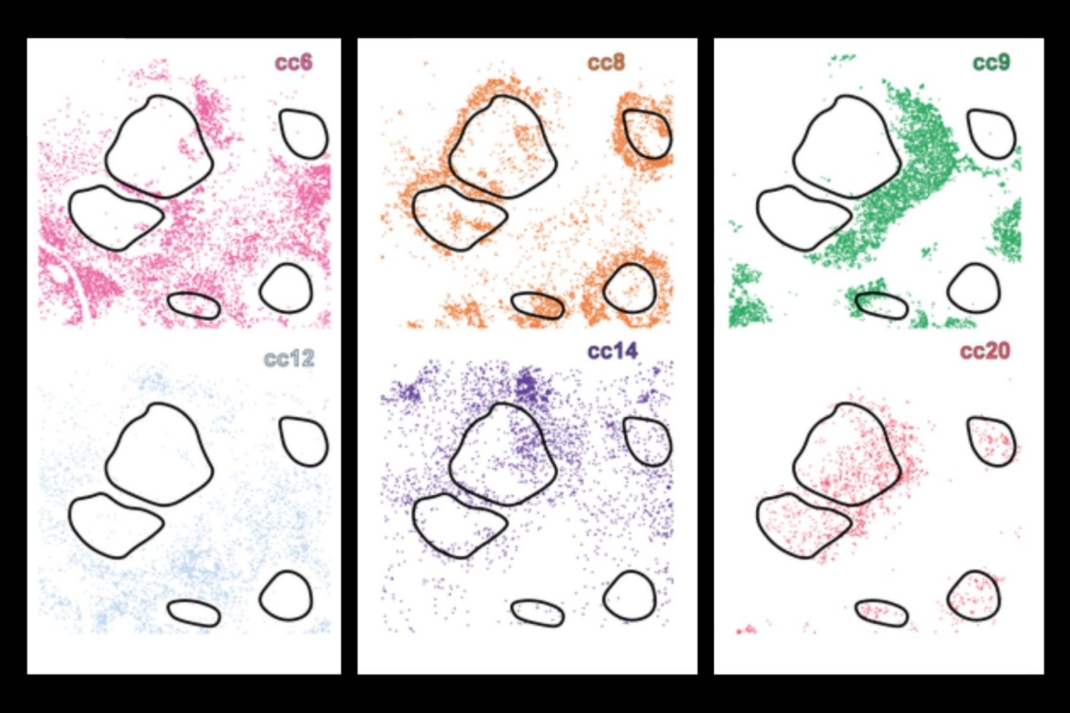
Historically, scientists have examined one or more of these facets in isolation, but now an innovative deep learning AI tool, CellLENS (Cell Local Environment and Neighborhood Scan), integrates all three dimensions, employing a blend of convolutional neural networks and graph neural networks to create a detailed digital representation for each individual cell. This functionality enables the system to categorize cells with analogous biology — effectively differentiating even those that seem very alike when viewed independently, yet behave distinctively based on their environment.
The research, recently published in Nature Immunology, outlines the findings of a partnership among researchers from MIT, Harvard Medical School, Yale University, Stanford University, and the University of Pennsylvania — spearheaded by Bokai Zhu, a postdoctoral researcher at MIT and a participant at the Broad Institute of MIT and Harvard as well as the Ragon Institute of MGH, MIT, and Harvard.
Zhu describes the significance of this new tool: “Initially, we might say, I discovered a cell. It is referred to as a T cell. By utilizing the same dataset through CellLENS, I can now assert that this is a T cell, and it is actively engaging a specific tumor boundary in a patient.
“I can apply existing knowledge to more accurately define what a cell is, its subpopulation, its actions, and the potential functional outputs of that cell. This approach could be leveraged to identify a new biomarker that provides particular and intricate details about diseased cells, facilitating the development of more targeted therapies.”
This marks a crucial advancement since current techniques frequently overlook vital molecular or contextual data — for instance, immunotherapies might focus on cells that exist solely at a tumor’s edge, restricting effectiveness. By employing deep learning, the researchers can uncover multiple layers of information with CellLENS, including cell morphology and spatial positioning within the tissue.
When applied to specimens from healthy tissues and various cancer types, including lymphoma and liver cancer, CellLENS revealed rare immune cell subtypes and demonstrated how their activity and positioning correlate with disease mechanisms — such as tumor infiltration or immune suppression.
These findings could aid scientists in gaining deeper insights into how the immune system interacts with tumors and set the stage for more precise cancer diagnostics and immunotherapies.
“I’m extremely enthusiastic about the promise of novel AI tools, like CellLENS, to enable us to more comprehensively understand abnormal cellular behaviors within tissues,” states co-author Alex K. Shalek, the director of the Institute for Medical Engineering and Science (IMES), the J. W. Kieckhefer Professor in IMES and Chemistry, and an extramural member of the Koch Institute for Integrative Cancer Research at MIT, along with being an Institute member of the Broad Institute and a member of the Ragon Institute. “We can now gather an immense quantity of information about individual cells and their tissue contexts using state-of-the-art, multi-omic assays. Effectively utilizing that data to propose new therapeutic options is an essential step in creating enhanced interventions. When combined with appropriate input data and meticulous downstream validations, such tools hold the potential to accelerate our capacity to positively influence human health and wellness.”
“`