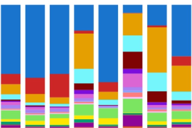
All existence is interconnected within an extensive familial framework. Each living entity relates to its predecessors, successors, and kin, and the lineage connecting any two beings can be traced. This is equally valid for cells within living organisms — every single one of the trillions of cells in the human body arises through continuous divisions from a fertilized egg, and all can be linked through a cellular lineage. In simpler life forms, such as the worm C. elegans, this cellular lineage has been completely charted; however, the cellular lineage of a human is significantly larger and more intricate.
Previously, MIT professor and Whitehead Institute for Biomedical Research member Jonathan Weissman, along with other scholars, devised lineage tracking techniques to monitor and reconstruct the genealogies of cell divisions in model organisms, aiming to deepen our understanding of the interrelations among cells and their assembly into tissues, organs, and, in certain instances, tumors. These techniques could provide insights into how organisms develop and how diseases like cancer begin and evolve.
Now, Weissman and his team have created a sophisticated lineage tracing instrument that not only accurately captures a genealogical tree of cell divisions but also integrates spatial information: determining the location of each cell within a tissue. The researchers utilized their tool, PEtracer, to examine the development of metastatic tumors in mice. By merging lineage tracing with spatial data, they gained a comprehensive perspective on how elements intrinsic to cancer cells and their surroundings influenced tumor development, as shared by Weissman and his postdocs Luke Koblan, Kathryn Yost, and Pu Zheng, along with graduate student William Colgan in a publication in the journal Science dated July 24.
“Creating this tool necessitated the amalgamation of varied skills through a type of ambitious interdisciplinary collaboration that’s achievable only at a place like Whitehead Institute,” remarks Weissman, who is also a Howard Hughes Medical Institute investigator. “Luke contributed expertise in genetic engineering, Pu in imaging, Katie in cancer biology, and William in computation, but the true key to their success was their capacity to collaborate in building PEtracer.”
“Comprehending how cells navigate through time and space is a vital approach to studying biology, and here we were capable of observing both aspects with high precision. The objective is to understand how various factors throughout a cell’s existence influenced its behaviors, by comprehending both its history and eventual position. In this research, we use these strategies to investigate tumor growth, but in principle, we can now begin to apply these tools for studying other biological phenomena of interest, such as embryonic development,” Koblan states.
Creating a tool to monitor cells in space and time
PEtracer tracks the lineages of cells by continuously appending short, predetermined sequences to their DNA over time. Each segment of code, referred to as a lineage tracing mark, consists of five bases, the fundamental components of DNA. These markers are introduced using a gene-editing technique called prime editing, which directly modifies sections of DNA with minimal unintended byproducts. Over time, each cell accumulates additional lineage tracing marks while preserving the marks of its ancestors. The researchers can thus juxtapose the combinations of marks among cells to deduce relationships and reconstruct the genealogy.
“We employed computational modeling to devise the tool from fundamental principles, ensuring its high accuracy and compatibility with imaging technology. We conducted numerous simulations to determine the optimal parameters for the new lineage tracing tool, and subsequently engineered our system to align with those parameters,” Colgan explains.
When the tissue — in this instance, a tumor developing in the lung of a mouse — had matured sufficiently, the researchers harvested these tissues and utilized advanced imaging methods to analyze each cell’s lineage relationship with other cells through the lineage tracing marks, along with its spatial position within the imaged tissue and its identity (as determined by the levels of various RNAs expressed in each cell). PEtracer is compatible with both imaging techniques and sequencing methods that capture genetic information from individual cells.
“Facilitating the collection and analysis of all this data from the imaging posed a significant challenge,” Zheng states. “What particularly excites me is not just that we managed to collect terabytes of data, but that we designed the project to gather information we knew would be pivotal in answering crucial questions and advancing biological discovery.”
Reconstructing a tumor’s history
By combining lineage tracing, gene expression, and spatial data, the researchers were able to comprehend how the tumor expanded. They could discern the closeness of neighboring cells and compare their characteristics. Utilizing this methodology, the researchers discovered that the tumors they examined comprised four distinct modules, or neighborhoods, of cells.
The tumor cells nearest to the lung, the most nutrient-rich region, exhibited the highest fitness, indicating their lineage history reflected the greatest rate of cell division over time. Fitness in cancer cells typically correlates with the aggressive growth of tumors.
The cells at the “leading edge” of the tumor, situated farthest from the lung, were more heterogeneous and less fit. Beneath the leading edge was a low-oxygen community of cells that may have once been at the leading edge, now confined in a less advantageous position. Nestled between these cells and those adjacent to the lung was the tumor core, a region containing both living and dead cells, along with cellular debris.
The researchers found that cancer cells across the family tree had an equal likelihood of being located in most regions, with the exception of the lung-adjacent region, where certain branches of the family tree were predominant. This indicates that the differing traits of cancer cells were significantly influenced by their environments or the conditions of their localized neighborhoods rather than their genealogical history. Further evidence supporting this notion was that the expression of particular fitness-related genes, such as Fgf1/Fgfbp1, correlated with a cell’s location rather than its ancestry. However, lung-adjacent cells also possessed inherited traits conferring advantages, such as the expression of the fitness-associated gene Cldn4 — demonstrating that ancestral history influenced outcomes as well.
These results illustrate how tumor development is shaped by both intrinsic lineage factors of specific cancer cells and by environmental elements that influence the behavior of the exposed cancer cells.
“By examining so many dimensions of the tumor simultaneously, we could derive insights that would not have been achievable with a more limited perspective,” Yost asserts. “Characterizing diverse populations of cells within a tumor will allow researchers to craft therapies that effectively target the most aggressive populations.”
“Now that we’ve accomplished the challenging task of designing the tool, we’re eager to apply it to investigate various questions in health and disease, in embryonic development, and across other model organisms, with the aim of addressing significant issues in human health,” Koblan adds. “The data we gather will also contribute to training AI models of cellular behavior. We look forward to sharing this technology with other researchers and discovering what we can all uncover.”

