“`html
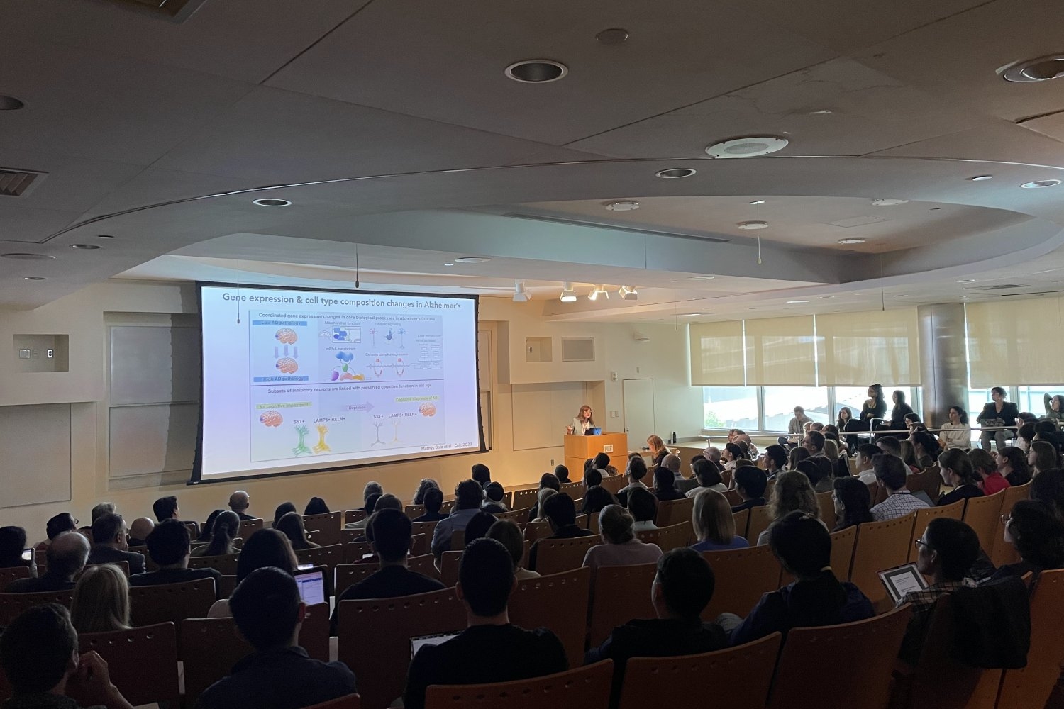
Grasping how connections between the central nervous system and the immune system influence aging issues, such as Alzheimer’s disease, Parkinson’s disease, arthritis, and more, can provide new avenues for therapeutic innovation, experts remarked at MIT’s symposium “The Neuro-Immune Axis and the Aging Brain” on Sept 18.
“The last ten years have seen swift advancements in our comprehension of how adaptive and innate immune systems affect the development of neurodegenerative conditions,” stated Picower Professor Li-Huei Tsai, head of The Picower Institute for Learning and Memory and MIT’s Aging Brain Initiative (ABI), during her introduction to the event, which had over 450 registrants. “Together, the speakers of today will delineate how the neuro-immune axis influences brain wellness and illness … Their research converges on the potential of immunology-informed interventions to slow or avert neurodegeneration and age-related cognitive decline.”
For example, keynote presenter Michal Schwartz from the Weizmann Institute in Israel elaborated on her years of groundbreaking research aimed at comprehending the neuro-immune “ecosystem.” Immune cells, she noted, assist in brain recovery and facilitate numerous functions, including its “plasticity,” or the capability to adjust to and absorb new information. However, Schwartz’s laboratory discovered that an immune signaling cascade can develop with age, which compromises cognitive abilities. She has utilized this knowledge to explore and develop remedial immunotherapies that enhance the brain’s immune reaction to Alzheimer’s by rejuvenating microglia immune cells and enlisting the support of peripheral immune cells called macrophages. Schwartz has advanced this prospective treatment to the market as the chief scientific officer of ImmunoBrain, a company trialing it in a clinical study.
In her address, Tsai mentioned recent findings from her lab and that of computer science professor and fellow ABI member Manolis Kellis indicating that many genes linked with Alzheimer’s disease are most prominently expressed in microglia, giving it a gene expression profile more akin to autoimmune disorders than to several psychiatric conditions (where expression of disease-related genes is typically highest in neurons). The investigation demonstrated that microglia become “exhausted” throughout disease advancement, losing their cellular identity and turning dangerously inflammatory.
“Genetic susceptibility, epigenomic instability, and microglia fatigue genuinely play a crucial role in Alzheimer’s disease,” Tsai remarked, adding that her lab is currently also exploring how immune T cells, recruited by microglia, may further influence the progression of Alzheimer’s disease.
The body and the brain
The neuro-immune “axis” not only links the nervous and immune systems but also extends throughout the entire body and the brain, with extensive implications for aging. Several presenters emphasized the primary pathway: the vagus nerve, connecting the brain to the body’s principal organs.
For instance, Sara Prescott, a researcher in the Picower Institute and an MIT assistant professor of biology, shared evidence her laboratory is gathering that indicates the brain’s signaling through vagus nerve terminals in the body’s airways is vital for managing the body’s defense against respiratory tissues. Considering that we breathe in approximately 20,000 times each day, our airways face various environmental challenges, Prescott noted, and her lab along with others is discovering that the nervous system interacts directly with immune pathways to generate physiological responses. However, vagal reflexes diminish with age, she pointed out, leading to increased vulnerability to infections; therefore, her lab is currently working with mouse models to analyze airway-to-brain neurons throughout the lifespan in order to better understand how they evolve with aging.
In his talk, Caltech Professor Sarkis Mazmanian concentrated on research in his lab connecting the gut microbiome to Parkinson’s disease (PD), particularly by promoting alpha-synuclein protein pathology and motor issues in mouse models. His lab theorizes that the microbiome can initiate alpha-synuclein in the gut via a bacterial amyloid protein that may subsequently encourage pathology in the brain, possibly through the vagus nerve. Based on its findings, the lab has devised two interventions. One involves administering a high-fiber diet to alpha-synuclein overexpressing mice to increase short-chain fatty acids in their gut, effectively modulating the activity of microglia in the brain. This high-fiber diet not only alleviates motor dysfunction but also corrects microglia activity and reduces protein pathology, he demonstrated. The other is a pharmaceutical agent aimed at disrupting the bacterial amyloid in the gut, preventing alpha-synuclein formation in the mouse brain and diminishing PD-like symptoms. These findings are awaiting publication.
Meanwhile, Kevin Tracey, a professor at Hofstra University and Northwell Health, took the audience on a journey along the vagus nerve to the spleen, explaining how impulses within the nerve regulate the immune system’s release of signaling molecules, or “cytokines.” An excessive surge can be detrimental, for example, leading to the autoimmune disorder rheumatoid arthritis. Tracey detailed how a newly U.S. Food and Drug Administration-approved pill-sized neck implant designed to stimulate the vagus nerve aids patients with severe forms of the disease without suppressing their immune system.
The brain’s border
Other presenters examined opportunities for comprehending neuro-immune interactions in aging and illness at the “boundaries” where the brain’s and body’s immune systems converge. These zones include the meninges surrounding the brain, the choroid plexus (located near the ventricles, or open areas, within the brain), and the interface between brain cells and the circulatory system.
For example, drawing from studies indicating that circadian disruptions can raise the risk for Alzheimer’s disease, Harvard Medical School Professor Beth Stevens from Boston Children’s Hospital shared new research from her lab that investigated how brain immune cells might behave differently concerning the day-night cycle. The project, spearheaded by newly minted PhD Helena Barr, discovered that “border-associated macrophages”—long-lasting immune cells situated in the brain’s borders —displayed circadian rhythms in gene expression and functioning. Stevens explained how these cells are calibrated by the circadian clock to “consume” more during the resting phase, a mechanism that may aid in removing debris draining from the brain, including Alzheimer’s disease-associated peptides like amyloid-beta. Therefore, Stevens hypothesizes that circadian disruptions, perhaps due to aging or night-shift employment, could contribute to disease onset by disturbing the fragile equilibrium in immune-mediated “clean-up” within the brain and its periphery.
Following Stevens, Washington University Professor Marco Colonna outlined how various categories of macrophages, including border macrophages and microglia, evolve from the embryonic stage. He elaborated on the distinct gene-expression programs that steer their differentiation into specific types. One gene he emphasized, for instance, is essential for border macrophages along the brain’s vasculature to assist in regulating the waste-clear cerebrospinal fluid (CSF) flow that Stevens also mentioned. Disabling the gene also hampers blood circulation. Importantly, his lab has identified that versions of the gene might offer some protection against Alzheimer’s, and that modulating the expression of the gene could represent a therapeutic approach.
Colonna’s WashU colleague Jonathan Kipnis (a former pupil of Schwartz) also discussed macrophages associated with the unique border separating brain tissue and the plumbing alongside the vasculature that transports CSF. His lab demonstrated in 2022 that these macrophages actively control the flow of CSF. He pointed out that eliminating the macrophages allowed the accumulation of Alzheimer’s proteins in mice. His lab continues to explore how these specific border macrophages might play roles in diseases. He is also examining separate studies on how the skull’s brain marrow contributes to the population of immune cells in the brain and may play a role in neurodegeneration.
Throughout discussions of distant organs and the brain’s borders, neurons themselves were never far from focus. Harvard Medical School Professor Isaac Chiu acknowledged their role in his presentation that concentrated on how they engage in their immune defense, for example, by directly detecting pathogens and emitting inflammation signals upon cellular death. He examined a crucial molecule involved in that latter process, which is expressed among neurons throughout the brain.
Whether scrutinizing within the brain, at its borders, or throughout the body, speakers demonstrated that age-related nervous system ailments are not only increasingly understood but also potentially better treated by considering not only the nerve cells but also their immune system collaborators.
“`

