Hazards appear, but they also vanish, and when they do, the brain receives an “all-clear” indication that educates it to diminish its trepidation. A recent investigation conducted on mice by MIT neuroscientists reveals that this indication is linked to the release of dopamine along a particular interregional brain circuit. The study thus highlights a potentially vital mechanism of mental wellness, reinstating tranquility when it functions properly, but extending apprehension or even post-traumatic stress disorder when it fails.
“Dopamine is crucial for commencing fear extinction,” states Michele Pignatelli di Spinazzola, co-author of the new research from the lab of senior author Susumu Tonegawa, Picower Professor of biology and neuroscience at the RIKEN-MIT Laboratory for Neural Circuit Genetics within The Picower Institute for Learning and Memory at MIT, and a Howard Hughes Medical Institute (HHMI) scholar.
In 2020, Tonegawa’s lab discovered that the process of learning to become fearful, and subsequently understanding when that fear is unnecessary, results from a rivalry among cell populations in the brain’s amygdala. When a mouse discovers that an area is “hazardous” (because it experiences a mild foot shock there), the fear memory is recorded by neurons in the anterior region of the basolateral amygdala (aBLA) expressing the gene Rspo2. When the mouse later finds that an area is no longer linked to peril (as it waits there and does not receive another shock), neurons in the posterior basolateral amygdala (pBLA) expressing the gene Ppp1r1b encode a new fear extinction memory that supersedes the original fear. Importantly, those same neurons also encode reward feelings, which helps clarify why it feels rewarding to realize that an expected threat has diminished.
In the current study, the lab, led by former members Xiangyu Zhang and Katelyn Flick, aimed to identify what stimulates these amygdala neurons to encode such memories. The comprehensive experiments the team reports in the Proceedings of the National Academy of Sciences demonstrate that it is dopamine being sent to the different amygdala populations from distinct clusters of neurons in the ventral tegmental area (VTA).
“Our research reveals a precise mechanism through which dopamine aids the brain in unlearning fear,” notes Zhang, who also led the 2020 study and is now a senior associate at Orbimed, a healthcare investment firm. “We discovered that dopamine stimulates specific amygdala neurons linked to reward, which consequently drive fear extinction. We now recognize that unlearning fear is not merely about suppressing it — it is a constructive learning process fueled by the brain’s reward system. This opens new pathways for understanding and perhaps treating fear-related disorders, such as PTSD.”
Forgetting fear
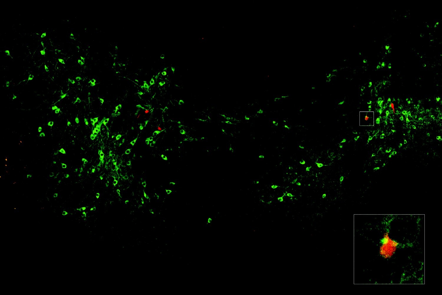
The VTA was the lab’s primary suspect for originating the signal, as this region is well known for encoding surprising experiences and guiding the brain, with dopamine, to learn from them. The initial set of experiments in the paper utilized various methods for tracing neural circuits to determine if and how cells in the VTA and amygdala interconnect. They identified a distinct pattern: Rspo2 neurons were influenced by dopaminergic neurons in the anterior and lateral aspects of the VTA. Ppp1r1b neurons received dopaminergic inputs from neurons in the central and posterior sections of the VTA. The density of connections was greater for the Ppp1r1b neurons than for the Rspo2 ones.
The circuit tracing demonstrated that dopamine is accessible to amygdala neurons involved in fear and its extinction, but do those neurons respond to dopamine? The team confirmed that indeed they express “D1” receptors for the neuromodulator. In line with the level of dopamine connectivity, Ppp1r1b cells had more receptors compared to Rspo2 neurons.
Dopamine fulfills numerous roles, so the subsequent question was whether its activity in the amygdala truly correlates with fear encoding and extinction. Using a method to track and visualize it in the brain, the team observed dopamine in the amygdala as mice underwent a three-day experiment. On Day One, they were placed in a chamber where they received three mild foot shocks. On Day Two, they returned to the chamber for 45 minutes without experiencing any additional shocks — initially, the mice froze in fear of another shock, but then relaxed after approximately 15 minutes. On Day Three, they returned once more to assess whether they had successfully extinguished the fear they exhibited at the beginning of Day Two.
The dopamine activity tracking illustrated that during the shocks on Day One, Rspo2 neurons exhibited a stronger response to dopamine. However, in the early stages of Day Two, when the anticipated shocks were absent and the mice began to relax, the Ppp1r1b neurons displayed heightened dopamine activity. More remarkably, the mice who successfully learned to extinguish their fear demonstrated the most substantial dopamine signal at those neurons.
Causal connections
The final sets of experiments aimed to demonstrate that dopamine is not merely available and associated with fear encoding and extinction, but also actively induces them. In one experiment, they utilized optogenetics, a technique allowing scientists to excite or suppress neurons using varying colors of light. Indeed, when they inhibited VTA dopaminergic inputs in the pBLA, this disruption hindered fear extinction. When the inputs were activated, it expedited fear extinction. The researchers were astonished that activating VTA dopaminergic inputs into the aBLA could restore fear even without any new foot shocks, undermining fear extinction.
The other method they employed to confirm a causal role for dopamine in fear encoding and extinction involved manipulating the dopamine receptors on amygdala neurons. In Ppp1r1b neurons, increasing dopamine receptor expression hindered fear recall and advanced extinction, while diminishing the receptors impaired fear extinction. Conversely, in the Rspo2 cells, reducing receptor levels diminished freezing behavior.
“We demonstrated that fear extinction necessitates VTA dopaminergic activity in the pBLA Ppp1r1b neurons through optogenetic inhibition of VTA terminals and cell-type-specific knockdown of D1 receptors in these neurons,” the authors noted.
The scientists cautiously highlight in the study that while they have identified the “teaching signal” for fear extinction learning, the overarching phenomenon of fear extinction occurs throughout the brain rather than in this single circuit.
Nonetheless, this circuit appears to be a critical point to examine as drug developers and psychologists strive to address anxiety and PTSD, Pignatelli di Spinazzola emphasizes.
“Fear learning and fear extinction offer a robust framework to explore generalized anxiety and PTSD,” he states. “Our study delves into the underlying mechanisms, suggesting multiple targets for a translational approach, such as pBLA and the utilization of dopaminergic modulation.”
Marianna Rizzo is also a co-author of the study. The research received support from the RIKEN Center for Brain Science, the HHMI, the Freedom Together Foundation, and The Picower Institute.

