
At the molecular and cellular scale, ketamine and dexmedetomidine function quite differently; however, in the surgical environment, they achieve the identical outcome: anesthetizing the patient. By illustrating how these varied substances attain the same result, a recent animal study conducted by neuroscientists at The Picower Institute for Learning and Memory at MIT highlights a potential mark of unconsciousness that can be easily measured to enhance anesthesiology practice.
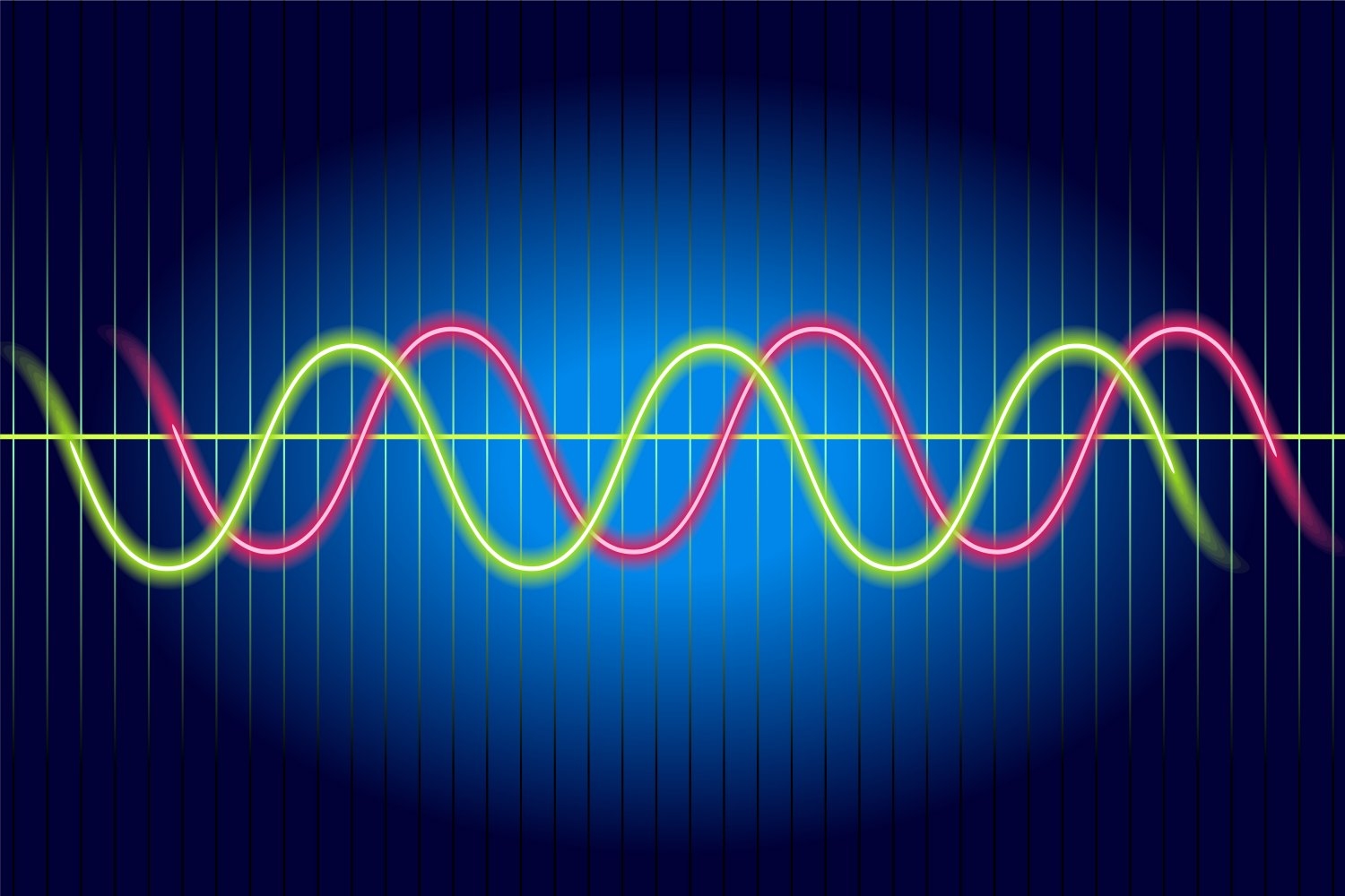
The commonality between the two drugs, the researchers found, lies in their influence on brain waves, which stem from the collective electrical activity of neurons. When brain waves align, meaning their peaks and troughs coincide, local clusters of neurons in the brain’s cortex can exchange information to generate conscious cognitive processes such as attention, perception, and reasoning, explains Picower Professor Earl K. Miller, the leading author of the new study published in Cell Reports. Conversely, when brain waves become misaligned, local communication and, hence, functions collapse, resulting in unconsciousness.
The discovery, spearheaded by graduate student Alexandra Bardon, not only enhances scientists’ comprehension of the boundary between consciousness and unconsciousness, Miller notes, but also could offer a unified new metric for anesthesiologists who utilize an array of different anesthetics to keep patients safely on the appropriate side of that boundary during surgical procedures.
“If you examine how the phase shifts in our recordings, you can hardly determine which drug was used,” states Miller, a faculty member at the Picower Institute and MIT’s Department of Brain and Cognitive Sciences. “That insight is crucial for medical practice. Furthermore, if unconsciousness features a universal signature, it could unveil the mechanisms that underlie consciousness.”
If more anesthetic agents also demonstrate similar effects on phase, then anesthesiologists might utilize brain wave phase alignment as a dependable indicator of unconsciousness as they adjust anesthetic drug dosages, Miller adds, irrespective of the specific combination of medications they are administering. This understanding could bolster efforts to create closed-loop systems that assist anesthesiologists by continually modifying drug dosages based on the brain wave readings of the patient’s state of unconsciousness.
Miller has been working with study co-author Emery N. Brown, an anesthesiologist and Edward Hood Taplin Professor of Computational Neuroscience and Medical Engineering at the Picower Institute, on developing such a system. In a recent clinical trial alongside colleagues in Japan, Brown demonstrated that monitoring brain wave power signals with EEG allowed an anesthesiologist to utilize significantly less sevoflurane during surgeries involving young children. The diminished doses were safe and correlated with numerous improved clinical outcomes, including a lesser occurrence of post-operative delirium.
Phase Discoveries
Neuroscientists examining anesthesia have seldom focused on phase; however, in the new research, Bardon, Brown, and Miller’s team emphasized it while anesthetizing two animals.
Following the animals’ loss of consciousness, the observations revealed a notable increase in “phase locking,” particularly at lower frequencies. Phase locking indicates that the relative phase differences became more stable. However, what captivated the researchers was the consistency that emerged: within each hemisphere, regardless of the anesthetic employed, brain wave phase became misaligned between the dorsolateral and ventrolateral areas of the prefrontal cortex.
Unexpectedly, brain wave phase across hemispheres became more synchronized, not less. Nevertheless, Miller notes that this is still a significant deviation from the conscious state, where brain hemispheres are usually not well-aligned, indicating that pronounced changes in phase alignment, although occurring in different manners at varying distances, correlate with unconsciousness relative to wakefulness.
“The increase in interhemispheric alignment of activity due to anesthetics seems to reverse the pattern observed in the awake, cognitively engaged brain,” the Bardon and Miller team wrote in Cell Reports.
Influenced by Distance
Distance emerged as a critical factor influencing the variation in phase alignment. Even across the 2.5 millimeters of a single electrode array, low-frequency waves exhibited a 20-30 degree misalignment. Over the approximately 20 millimeters separating arrays in the upper (dorsolateral) and lower (ventrolateral) regions within a hemisphere, this implies an approximate 180-degree shift in phase alignment, resulting in a complete offset of the waves.
This reliance on distance aligns with the concept of waves propagating across the cortex, Miller states. Indeed, in a 2022 study, Miller and Brown’s labs illustrated that the anesthetic propofol induced a strong low-frequency traveling wave that swept directly across the cortex, overpowering higher-frequency linear and rotational waves.
The new findings present numerous opportunities for future investigations, Miller asserts. Does propofol also create this signature of altered phase alignment? What role do traveling waves play in this phenomenon? Moreover, considering that sleep is also marked by heightened power in slow wave frequencies but is certainly distinct from anesthesia-induced unconsciousness, could phase alignment elucidate the variance?
In addition to Bardon, Brown, and Miller, the paper includes contributions from authors Jesus Ballesteros, Scott Brincat, Jefferson Roy, Meredith Mahnke, and Yumiko Ishizawa.
Support for the research was provided by the U.S. Department of Energy, the National Institutes of Health, the Simons Center for the Social Brain, the Freedom Together Foundation, and the Picower Institute.