
When an individual experiences a trauma to the spinal cord, the usual interactions between the brain and the spinal pathways below the injury get disrupted, leading to paralysis. As the brain continues to operate normally, along with the spinal cord beneath the injury, scientists have been striving to re-establish the communication to facilitate rehabilitation and possibly recover movement.
Researchers headed by Ismael Seáñez have designed a specific decoder to reinstate that communication. Seáñez serves as an assistant professor of biomedical engineering at the McKelvey School of Engineering, along with a position in neurosurgery at the School of Medicine, both situated at Washington University in St. Louis. His laboratory comprises doctoral student Carolyn Atkinson. Through investigations in their lab involving 17 human participants without a spinal cord injury, they successfully prompted movement in the lower leg using transcutaneous spinal cord stimulation, which involves noninvasive, external electrical pulses.
The findings of the study were published online on April 25 in the Journal of NeuroEngineering and Rehabilitation.
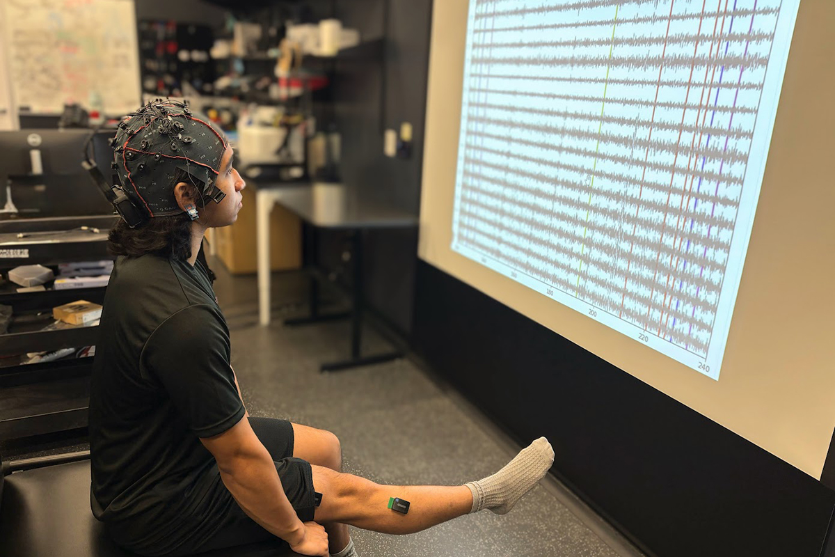
The team utilized a unique cap equipped with noninvasive electrodes that detect brain activity via electroencephalography (EEG). While wearing this cap, seated volunteers were instructed to extend their leg at the knee, and then merely to visualize extending their leg — while keeping it motionless — allowing researchers to capture the brain waves during both tasks.
The team supplied the neural activity data to the decoder, or algorithm, enabling it to comprehend how the brain waves function under both circumstances. They discovered that the genuine movement and imagined movement employed similar neural tactics.
“Once we provide the decoder with this information, it learns to anticipate based on neural activity whenever movement is or isn’t taking place,” Seáñez stated. “We demonstrate that we can anticipate when someone is contemplating moving their leg, even if their leg does not genuinely shift.”
The team implemented controls to guarantee that the participants were authentically imagining movement.
“Anytime individuals are in motion, this can introduce signal noise, and we aim to ensure that the signal noise is not what we’re learning to anticipate,” Seáñez remarked. “It’s the intention to move or the neural activity we want to predict, thus we have individuals visualize extending their leg and apply the identical algorithm trained on moving subjects to determine whether they were merely imagining or not.”
Seáñez noted that this uncovers two insights.
“Firstly, it’s more probable that we’re decoding movement intention rather than an artifact or noise, and secondly, when we implement this on individuals with spinal cord injuries who cannot genuinely move their legs for us to classify the data, we could use their imagination of moving a leg to educate our decoder.”
Seáñez stated that this proof-of-concept study marks an initial phase towards creating a noninvasive brain-spine interface that utilizes real-time predictions to deliver transcutaneous spinal cord stimulation, reinforcing voluntary movement in a single joint during rehabilitation for spinal cord injury patients.
Moving forward, the team intends to evaluate a generalized decoder trained on data from all participants to ascertain whether a universal decoder could operate as effectively as a personalized one, simplifying its application in clinical environments.
Atkinson C, Lombardi L, Lang M, Keesey R, Hawthorn R, Seitz Z, Leuthardt EC, Brunner P, Seáñez I. Development and evaluation of a non-invasive brain-spine interface using transcutaneous spinal cord stimulation. Journal of NeuroEngineering and Rehabilitation, online April 25, 2025. DOI: https://doi.org/10.1186/s12984-025-01628-6.
Funding for this research was provided by the McDonnell Center for Systems Neuroscience at Washington University in St. Louis; the National Institutes of Health (K12-HD073945, K01-NS127936; R01-EB026439; P41-EB018783); the Department of Biomedical Engineering in McKelvey Engineering at WashU; and the Department of Neurosurgery at WashU Medicine.
Originally published on the McKelvey Engineering website
The post Brain decoder controls spinal cord stimulation appeared first on The Source.