“`html
A microneedle patch detects cancer biomarkers in the outermost layer of skin to identify melanoma in animal tissue specimens
Research conducted by the University of Michigan suggests that melanoma testing could potentially be performed at home using a skin patch and a test strip featuring two lines, resembling at-home COVID-19 tests.
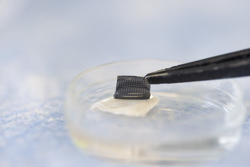
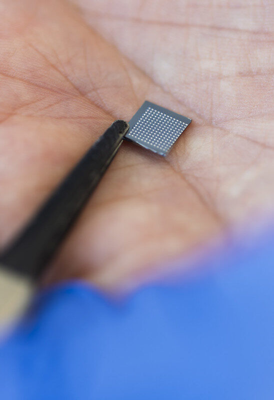
Funded by the National Institutes of Health, the innovative silicone patch with star-shaped microneedles, named the ExoPatch, successfully identified melanoma from healthy skin in mice.
This patch and testing mechanism pave the way for rapid at-home melanoma testing, enabling individuals to detect the most aggressive type of skin cancer early without needing a biopsy or blood sample.

“The star-shaped microneedles make the puncturing process easier and less painful; they are so diminutive that they only penetrate the outermost skin layer, the epidermis, without drawing any blood,” explained Sunitha Nagrath, the Dwight F. Benton Professor of Chemical Engineering at U-M and co-corresponding author of the research published in Biosensors and Bioelectronics.
The ExoPatch microneedles, measuring just 0.6 mm in length and with a tip width of less than 100 nanometers (0.0001 mm), are coated with a gel that captures exosomes—tiny vesicles released by cells—from the interstitial fluid that fills the gaps between cells within the epidermis.
Previously considered waste expelled from cells for disposal, exosomes are now understood to contain DNA and RNA fragments that facilitate communication between cells. Exosomes from cancer cells can help tumors spread by conditioning tissues to accept tumor cells ahead of their arrival, and identifying them can enable earlier cancer detection compared to earlier methodologies.
The gel enveloping the ExoPatch incorporates a protein known as Annexin V, which attracts and binds exosomes to the surface of the microneedles. After removal from the skin, immersing the patch in an acidic solution dissolves the gel, releasing the exosomes into a liquid medium. By dipping a test strip into this solution, two lines appear if melanoma exosomes are present, while a single line indicates a negative result—similar to how an at-home COVID-19 test strip operates.
“Individuals with fair skin and moles typically must visit their doctor every six months to send a biopsy for determining whether they are malignant or benign. With this testing method, they could, instead, test at home, receive immediate results, and follow up with a dermatologist if the result is positive,” stated Nagrath.
In an initial stage of the proof-of-concept study, the team examined the ExoPatch on a tissue sample of pig skin, which closely mirrors human skin regarding thickness and composition. Using microscope analysis, they noted that the microneedles penetrated approximately 350 to 600 nanometers into the skin. For reference, the epidermis on a human forearm typically measures around 18,300 nanometers thick.
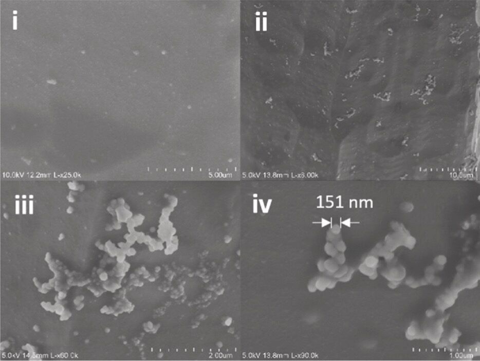
To evaluate whether the ExoPatch could capture melanoma exosomes from skin tissue, the research team analyzed tissue samples from mouse skin—half sourced from healthy mice and half from mice injected with a segment of a human melanoma tumor. Following a 15-minute application, the ExoPatch was examined under a high-powered microscope.
“Upon reviewing the microscopy images, I was pleased to see how effectively the exosomes attached to the microneedles and fell within the anticipated size range of 30 to 150 nanometers,” remarked Scott Smith, a doctoral candidate in chemical engineering at U-M and co-lead author of the study.
“““html Upon verifying that the exosomes adhered to the ExoPatch, the scientists dissolved the gel and processed the specimen through the test strips. The assay effectively differentiated between melanoma and healthy tissues, with a 3.5-fold darker line evident in melanoma samples. The ExoPatch extracted 11.5 times more exosomal protein from melanoma tissue specimens compared to healthy tissues, indicating its capability to specifically hone in on cancerous exosomes. A preliminary study in humans, followed by a series of clinical evaluations, will be the subsequent steps to advance this technology for practical application. Moreover, the ExoPatch gel coating could be adapted to identify exosomes released by various cancers with solid tumors, including lung, breast, colon, prostate, and brain cancers. “This is the inaugural patch designed to collect disease-specific exosomes from interstitial fluid beneath the skin. The potential uses are extensive,” explained Nagrath. This research is supported by the National Institutes of Health (1-R01-CA-208335-01-A1). The device was developed at the Lurie Nanofabrication Facility, and evaluations were partially conducted within the Biomedical Research Core Facilities and Rogel Cancer Center Immunology Core. The team has sought patent protection with the aid of U-M Innovation Partnerships. “`