
“`html
A Caltech-led group has created a secure, effective, and non-invasive breast imaging method that utilizes machine learning to assist in distinguishing between abnormal and healthy tissue. This approach has been evaluated on patients and exhibits performance that is equal to or superior to other standard breast imaging methods.
For many years, X-ray mammography has been regarded as the benchmark for breast imaging aimed at the early identification of breast cancer. While this technique continues to provide significant benefits in reducing cancer-related fatalities, it subjects patients to minor exposures of ionizing radiation, causes discomfort due to the tight compression of breasts to facilitate X-ray passage, and often leads to numerous false positive diagnoses, particularly in cases of dense breast tissue.
Alternative modalities such as ultrasound and magnetic resonance imaging (MRI) can also be employed for breast imaging, but they come with their own issues. Ultrasound is quite safe; however, its precision relies heavily on the operator’s skill, and the outcomes are not always definitive. MRI is labor-intensive, costly, and is not suitable for patients with allergies to contrast agents, those who experience claustrophobia, or individuals with certain implants.
“We were immensely driven to address this issue because none of the existing methods are flawless,” states Lihong Wang, the Bren Professor of Medical Engineering and Electrical Engineering at Caltech. “The future of healthcare must be superior to this.”
The method developed and enhanced by Wang and his team over the last two decades is termed photoacoustic computed tomography, or PACT. It presents a breast imaging alternative that eliminates the discomfort, high costs, or dangers linked with traditional evaluation techniques. PACT features a laser-sonic scanner capable of detecting tumors in as few as 15 seconds.
Collaborating with scientists at the City of Hope Comprehensive Cancer Center in Duarte, California, the team has assessed PACT on 39 patients. It achieved results comparable to mammography and MRI in distinguishing between suspicious and typical tissue as well as malignant and benign masses.
The researchers elaborate on PACT and their clinical findings in a new article published in the journal Nature Biomedical Engineering. The principal authors of the paper include Xin Tong (MS ’21), Cindy Z. Liu, and Yilin Luo, graduate students in the Andrew and Peggy Cherng Department of Medical Engineering at Caltech; along with Li Lin (PhD ’20), who completed the work while at Caltech and is currently at Zhejiang University in China.
“This is the culmination of literally decades of effort,” remarks Wang, who also holds the Andrew and Peggy Cherng Medical Engineering Leadership Chair and serves as the executive officer for medical engineering at Caltech. “Our goal is to develop PACT into a clinical tool that serves patients—enabling the detection of breast cancer without exposing them to the risks of cancer or concerns regarding allergic reactions.”
How It Operates
PACT functions by directing a near-infrared laser pulse into breast tissue. The laser light disperses throughout the breast and is absorbed by various molecules. For instance, it can be absorbed by hemoglobin molecules in the patient’s red blood cells, which results in the molecules vibrating ultrasonically.
Unlike X-rays that flow in a linear path, light waves scatter or bounce within tissues, complicating the acquisition of high-resolution images. Thus, PACT merges light and sound into a single technique. “We utilize light to visualize the molecules, but sound helps us define their spatial location,” explains Wang.
The vibrations emitted by the molecules traverse the tissue and are detected by an array of 512 small ultrasonic sensors placed over the skin of the breast. The data from these sensors are processed to create an internal image of the breast’s structures, similar to ultrasound imaging but with significantly greater precision. PACT can illustrate features as small as a quarter of a millimeter at a depth of 4 centimeters.
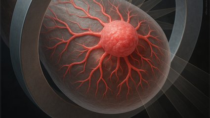
“We essentially use molecules to understand the body’s physiology,” remarks Wang. “That is the advantage of photoacoustic tomography: By identifying molecules, we can ascertain precisely how the body is functioning. When there is a functional variation, it indicates that we can potentially identify diseases more effectively.” For example, PACT excels at detecting hemoglobin, which reveals angiogenesis, a typical indicator of breast cancer involving the formation of new blood vessels to supply additional nutrient-rich blood to cancer cells. PACT can also identify tumor hypoxia, another cancer marker, where rapid metabolism exceeds blood supply, causing certain tumor regions to be deprived of oxygen.
Machine Learning Can Identify Suspicious Tissue, Sometimes Before Humans Can
With the advancements in artificial intelligence (AI) and machine learning, Wang asserts that PACT has become more adept at identifying irregularities in breast tissue than in prior years. The team trained the system using images of malignant and benign masses, along with suspicious and healthy tissue, thereby enhancing its capability to detect subtle differences that indicate the type of imaged tissue. Indeed, Wang indicates that PACT can frequently recognize problematic features that might go unnoticed by the human eye.
The Patient Experience
During a PACT examination, the patient lies face down on a table that contains a warm water bath, ultrasonic sensors, and the laser. One breast is positioned in the recess, and the laser shines into it from underneath. Due to the rapid nature of the technique, each scan is completed while the patient holds their breath.
“We began with a basic laboratory setup—a single-element ultrasound transducer that we rotated around—and it was a lengthy process. Now we can achieve 3D imaging within a single breath, making it very feasible,” remarks Wang.
Wang adds that in many respects, the new work is merely the beginning, as the team believes it can further enhance the imaging quality by incorporating additional laser wavelengths beyond the two they currently employ and refining other features. Looking ahead, the team’s forthcoming steps include gathering a more extensive dataset from additional breast cancer volunteers, refining the classification model by leveraging more features, and ultimately commercializing the technology.
The article is titled “Panoramic photoacoustic computed tomography with learning-based classification enhances breast lesion characterization.” Additional contributors include Peng Hu (PhD ’23) and Caltech graduate student Junfu Zheng; former Wang lab members Yide Zhang and Rui Cao; and Jessica Dzubnar, Marta Invernizzi, Stephanie Delos Santos, Jaclene Torres, Armine Kasabyan, Lily Lai, and Lisa D. Yee of City of Hope. This research was partially supported by grants from the National Institutes of Health.
“`