

“`html
An innovative design enables a method for examining metals, ceramics, and stones in a typical laboratory, broadening accessibility for students, academic researchers, and industry
For the first time, scientists can examine the microstructures within metals, ceramics, and rocks using X-rays in an ordinary lab without the need to visit a particle accelerator, as indicated by a study led by engineers from the University of Michigan.
This innovative approach renders 3D X-ray diffraction—referred to as 3DXRD—more accessible, potentially facilitating rapid evaluation of samples and prototypes in academic and industrial settings, while enhancing opportunities for students.
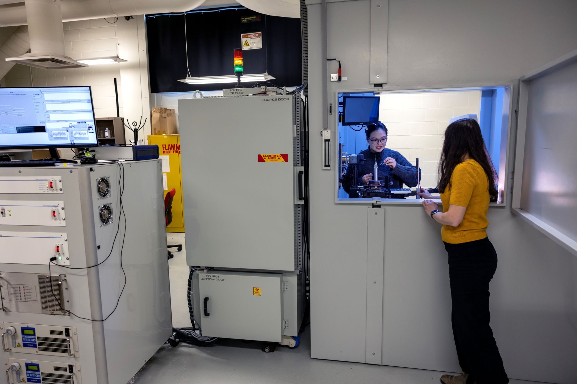
3DXRD generates three-dimensional images by capturing X-rays from various angles, akin to a CT scan. Rather than the imaging device revolving around a subject, a small material sample, just a few millimeters in width, rotates on a platform in front of a robust beam that emits about a million times more X-rays than a conventional medical X-ray.
The substantial concentration of X-rays creates a microscope-scale image of the minute fused crystals that comprise most metals, ceramics, and stones—referred to as polycrystalline materials.
The findings assist researchers in deciphering how materials respond to mechanical stresses by analyzing the volume, position, orientation, and strain of thousands of individual crystals. For instance, imaging a sample from a steel beam under pressure can reveal how the crystals manage the load of a building, aiding researchers in understanding large-scale wear.

Previously, synchrotrons were the sole facilities capable of producing sufficient X-rays for 3DXRD, as electrons emit a large number of X-rays while traversing through circular particle accelerators, which can subsequently be directed onto a sample.
Although synchrotron X-ray beams deliver cutting-edge detail, there are approximately only 70 such facilities globally. Research teams must submit proposals for “beam time,” and accepted projects often face wait times extending from six months to two years to execute their experiments, which are restricted to a maximum duration of six days.
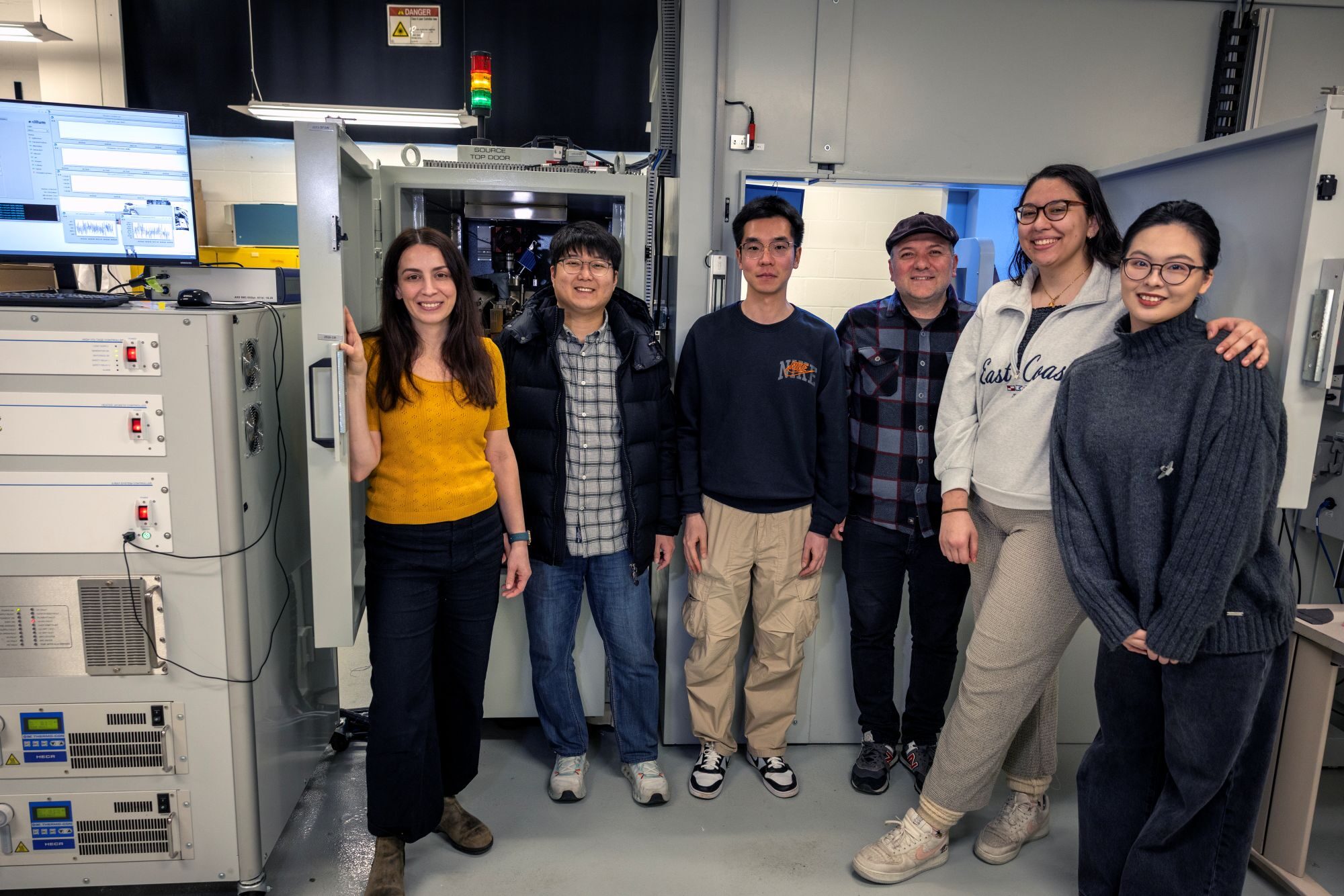
In a bid to render this technique more broadly accessible, the research team collaborated with PROTO Manufacturing to custom-create the inaugural laboratory-scale 3DXRD. In total, the instrument is comparable in size to a typical residential bathroom, but it could be miniaturized to fit within a closet space.

“This method provides such fascinating data that I aimed to establish the opportunity to explore new, high-risk, high-reward endeavors, allowing teachable moments for students without the delays and pressures associated with synchrotron beam time,” stated Ashley Bucsek, assistant professor of mechanical engineering and materials science and engineering at U-M, and co-corresponding author of the study published in Nature Communications.
Historically, small-scale apparatuses could not generate enough X-rays for 3DXRD, as at a certain threshold, the electron beam feeds excessive power into the anode—the solid metal surface that the electrons impact to create X-rays—resulting in melting. Lab-3DXRD employs a liquid-metal-jet anode that remains liquid at room temperature, enabling it to absorb more power and produce greater quantities of X-rays than previously feasible at this scale.
The researchers verified the design by scanning the identical titanium alloy sample through three approaches: lab-3DXRD, synchrotron-3DXRD, and laboratory diffraction contrast tomography or LabDCT—a method utilized to chart crystal structures in 3D, devoid of strain data.
Lab-3DXRD exhibited high precision, with 96% of the crystals it detected coinciding with those picked up by the other two methods. It performed particularly well with larger crystals exceeding 60 micrometers, though it overlooked some smaller crystals. The researchers mention that incorporating a more sensitive photon-counting detector, which detects X-rays used to construct the images, might aid in capturing finer-grained crystals.
With this technique now available on-site, Bucsek’s research group can explore new experiments, fine-tuning parameters in preparation for larger trials at a synchrotron.
“Lab-3DXRD is analogous to a nice backyard telescope while synchrotron-3DXRD resembles the Hubble Telescope. There are still scenarios where you require the Hubble, but we are now well-prepared for those major experiments because we can conduct preliminary tests,” Bucsek remarked.
In addition to making experiments more accessible, lab-3DXRD empowers researchers to extend projects beyond the six-day limit imposed at synchrotrons, which is particularly advantageous for studying cyclic loading—how a material reacts to repeated stresses over numerous cycles.
The first author and co-corresponding author Seunghee Oh, a research fellow in mechanical engineering at the time of the study, currently works in the X-ray Science Division at Argonne National Laboratory.
This research has been supported by the National Science Foundation (CMMI-2142302; DMR-1829070) and the U.S. Department of Energy (Award DE-SC0008637).
Researchers from PROTO Manufacturing also played a role in this study.
LabDCT was conducted at the Michigan Center for Materials Characterization.
“`