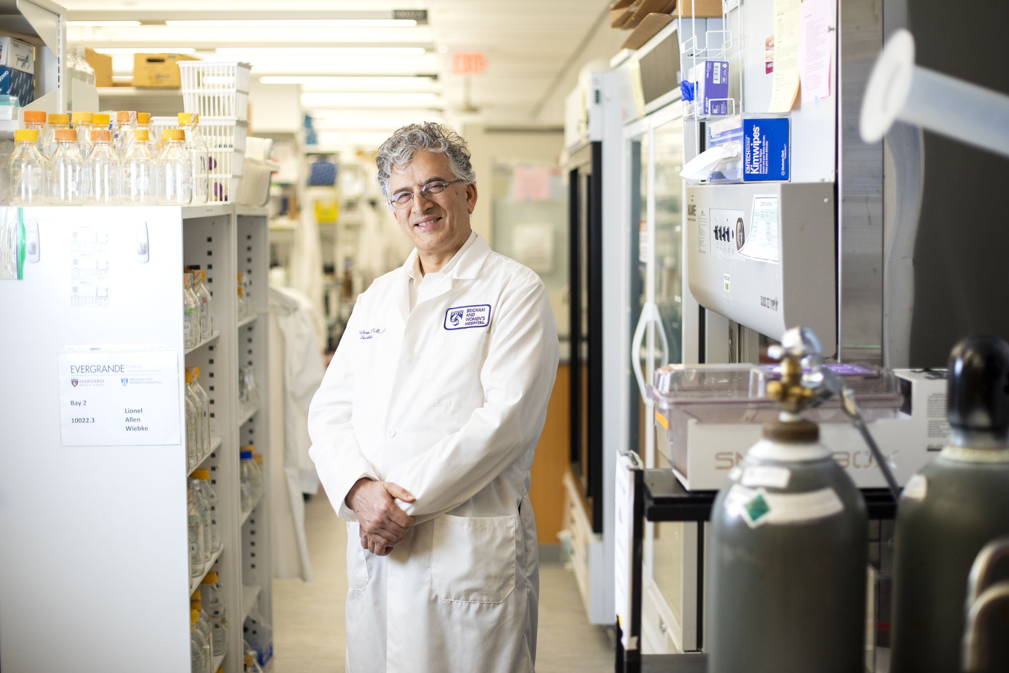


Vijay Kuchroo.
Veasey Conway/Harvard Staff Photographer
Health
Immune-system approach utilized to combat cancer may assist with Alzheimer’s
Turning off checkpoint molecules enabled microglia to confront plaques in the brain, enhancing memory in rodents
A recent study increases the likelihood that a technique already effective against certain cancers could be applied against Alzheimer’s. The investigation, which underscores the function of an immune system “checkpoint” molecule, demonstrated enhanced cognitive abilities in trials with mice. It was published earlier this month in Nature.
In this edited dialogue, the Gazette engaged with Vijay Kuchroo, the Samuel L. Wasserstrom Professor of Neurology at Harvard Medical School and Brigham and Women’s Hospital, and head of the Gene Lay Institute of Immunology and Inflammation at Brigham and Women’s Hospital, Massachusetts General Hospital, and Harvard Medical School.
Kuchroo, who served as a senior author on the article, elaborated on research that eliminated the expression of a molecule known as TIM-3, which prevents brain immune cells referred to as microglia from combatting Alzheimer’s plaques, thereby freeing these cells to eliminate plaques and restore memory.
Your research focused on a model of late-onset Alzheimer’s disease. How many cases does this represent?
Most instances of Alzheimer’s disease (AD), between 90 percent and 95 percent, fall under late-onset. The molecule we investigated, known as TIM-3, was associated with late-onset Alzheimer’s through a genome-wide association study and identified as a genetic risk factor for the condition. A polymorphism in the TIM-3 gene, HAVCR2, exists in AD patients. TIM-3 is an inhibitory molecule used by the immune system to deactivate immune cells once they are activated. TIM-3 is part of a category of inhibitory molecules termed checkpoint molecules, which have been utilized in cancer treatments.
Checkpoint molecules prevent the body from attacking its own tissues?
That’s one way to describe it. When your immune system is activated, checkpoint molecules help to restrain the immune response from becoming excessive.
An excellent illustration is when you have an infection such as the common cold; your lymph nodes swell as millions of T cells are produced to fight the virus. Once the infection subsides, checkpoint molecules come in to reduce the number of T cells to their normal state.
Cancers have taken advantage of these checkpoint molecules for survival, as every time a T cell attempts to assault a tumor cell, the tumor cell induces the expression of checkpoint molecules, preventing the T cells from attacking. As a result, T cells become dysfunctional or exhausted, allowing the tumor to persist.
The new angle is that in Alzheimer’s disease, there is a build-up of plaques in the brain that macrophage-like cells known as microglia fail to clear. These microglia show increased expression of the checkpoint molecule TIM-3.
They essentially act as the immune cells within the brain?
Microglia function as the immune cells of the brain and carry out other vital roles. During development, synapses are formed, which are crucial for memory storage. The issue arises as even fleeting experiences can create memories, necessitating the removal of memories that are no longer useful. Hence, the primary role of microglia during development is to prune infrequently used synapses to refine and maintain memory.
Once you are born and have established memories, it’s undesirable to lose them. Thus, around 28 to 40 days post-birth in mice, and a few months to years in humans, there is a developmental mechanism causing microglia to cease pruning in order to retain formed memories.
To halt microglial pruning, these cells increase their expression of the checkpoint molecule TIM-3, rendering microglia homeostatic and preventing them from phagocytosing any longer.
That is advantageous because you do not want to prune your own memories, yet it produces a downside as one ages, leading to a build-up of debris in the brain that remains unaddressed. Who will clean it up? Microglia become homeostatic and TIM-3 prevents them from engulfing the accumulated debris, leading to plaque formation.
What distinguishes TIM-3 in an older individual with Alzheimer’s compared to one without it?
A polymorphism is present in the gene, and Alzheimer’s patients with this polymorphism exhibit significantly heightened TIM-3 expression on microglia versus those without the disease.
So, all that TIM-3 keeps the microglial cells at equilibrium and prevents them from attacking amyloid beta plaques even though they are detrimental to the brain?
Indeed, microglia should be responsible for clearing amyloid plaques, yet they do not fulfill this role. We identified this molecule on T cells in the immune system, but it is present at a level 100 times — and in some cases 1,000 times — greater on activated microglia.
Thus, the same molecule that restores T cell population to normal after an infection is employed by microglia to prevent excessive pruning. However, it also becomes a liability as it obstructs them from attacking the plaques that build up in Alzheimer’s disease.
You experimented with lab mice that lack the HAVCR2 gene — responsible for making TIM-3?
Yes, these mice were engineered to investigate TIM-3’s role in immune system autoimmunity and cancer.
We utilized the same mice. We genetically knocked out the gene, leading to microglia that do not express TIM-3 upon activation. This boosts the clearance of plaques and alters their behavior.
Toxic plaques have fingerlike protrusions that invade the brain, but as microglia nibble on them, the plaques become more compact. Therefore, eliminating TIM-3 in microglia not only reduces plaque quantity but also enhances the quality of the plaque. These mice actually regain some cognition. Not entirely, but there is a notable improvement in their cognitive behavior.
When we refer to assessing cognitive behavior in mice, do we mean their capacity to recall and maneuver through mazes?
That is accurate. Mice burdened with plaques in their brains tend to remember less. They also exhibit diminished fear responses. In an open area, normal mice will retreat to a corner to avoid becoming prey. However, when burdened with plaques, they will remain in the center of the maze, lacking defensive instincts. Once the plaques are removed, memory returns, and their fear responses normalize since having an appropriate level of fear is crucial for survival.
What would a TIM-3 therapy entail for human patients with Alzheimer’s?
The therapy would involve using an anti-TIM-3 antibody or a small molecule capable of inhibiting TIM-3’s suppressive function.
What is the potential impact of this approach on Alzheimer’s disease? After numerous unsuccessful drug trials, recent studies have yielded some improvements, albeit minor.
As amyloid beta is found in endothelial cells within blood vessels, a large volume of antibodies does not reach the brain as they attack the blood vessels, leading to strokes due to vascular damage, thus limiting the use of anti-amyloid antibodies in AD treatment. Given TIM-3’s selective expression, existing anti-TIM-3 antibodies can be repurposed for Alzheimer’s treatment.
How long did this research take?
Five years; each experiment typically took around eight to nine months. I would like to highlight that this was a collaboration with my colleague Oleg Butovsky at the Ann Romney Center for Neurological Disease. Approximately six individuals, three from my lab and three from his lab, worked diligently to conduct these experiments.
What are the next steps?
We are investigating whether human anti-TIM-3 can prevent plaque formation in Alzheimer’s disease mouse models. We have created a mouse model in which the human TIM-3 gene has been incorporated, making it ideal for testing various candidate antibodies for human applications.
This study was partially funded by the National Institutes of Health.