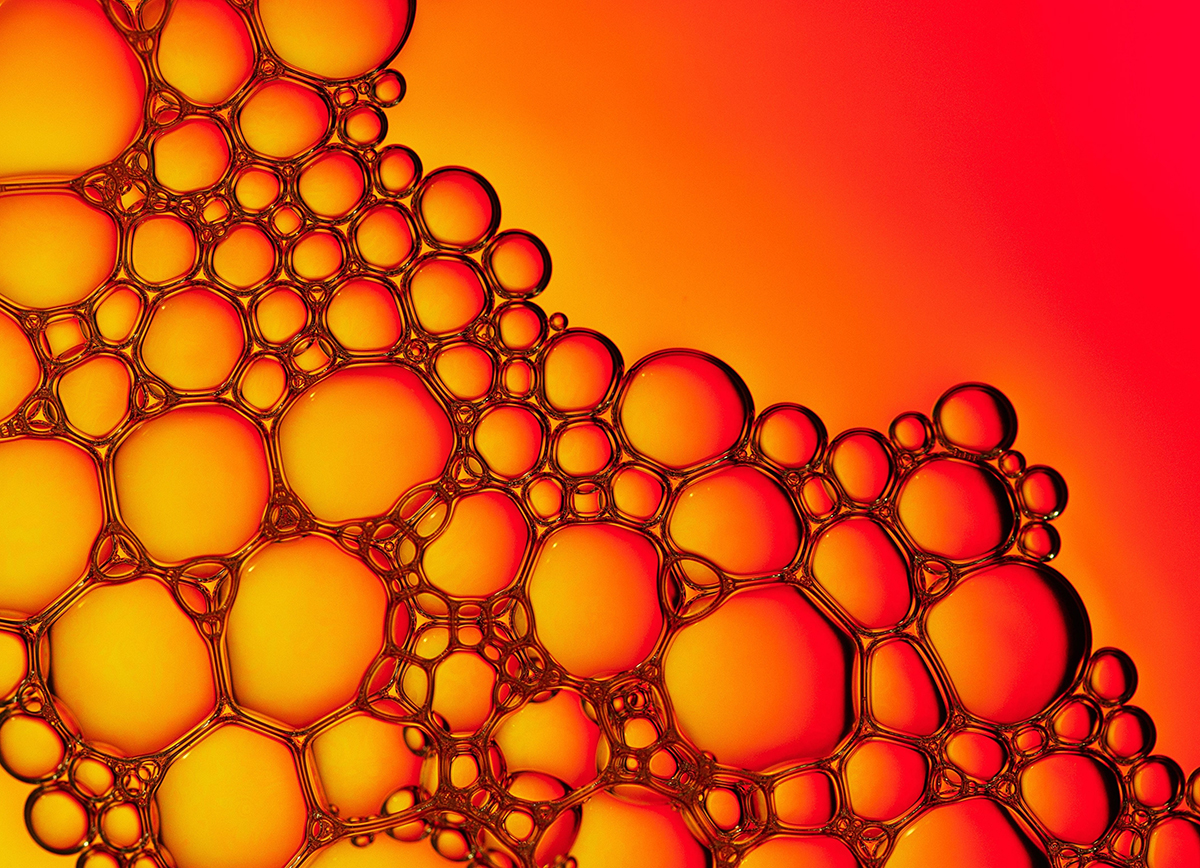
Biomolecular condensates are dynamic entities within our cells that orchestrate cellular components. They represent unique molecular collectives composed of DNA, RNA, and proteins that “condense” molecules to specific sites, yet they often challenge conventional descriptions. This partly arises from their diminutive size, which eludes measurement by standard microscopes.
“These entities were previously characterized as ‘liquid-like’ because some were seen to coalesce, merge, drip, and flow similar to raindrops on car windows,” remarked Rohit Pappu, the Gene K. Beare Distinguished Professor of biomedical engineering at the McKelvey School of Engineering, Washington University in St. Louis.
Nonetheless, despite their raindrop-like appearance, computational analyses suggest different behavior. The molecular arrangement in condensates resembles a network that reorganizes across various time frames, imparting a more dynamic, Silly Putty-like nature to them.
Collaborating with Matthew Lew’s lab, an associate professor of electrical and systems engineering at McKelvey Engineering, Pappu and his team, all part of the WashU Center for Biomolecular Condensates, examined computational forecasts by investigating condensates with innovative super-resolution microscopy techniques.
In research released on March 14 in Nature Physics, the Pappu and Lew laboratories demonstrate how to utilize fluorogens — dyes sensitive to environmental changes that illuminate only in specific chemical conditions — to explore condensates at high resolutions previously unattainable by scientists. They employed these fluorogens individually, supported by specialized imaging methods developed in the Lew lab.
These advancements in imaging are pivotal for comprehending the mechanisms of condensates and how they malfunction in relation to cancers and neurodegenerative disorders, which are linked to dysfunctional condensates.
Current methodologies depend on averaging the behavior of the entire ensemble of molecules within condensates. Conversely, this novel single fluorogen technique facilitates single and multimolecular resolution. The WashU team utilized fluorescent chemical markers that illuminate only when they interact with the appropriate type of chemical and viscoelastic environment, acting as signals for areas that appear as hubs within networks of molecules held together by “stickers.”
Regarding those stickers: If you visualize condensates as a gathering of individuals, the stickers represent the acquaintances who determine where and when to assemble and whom to include, according to the researchers.
“Powered by the interactions embedded within protein sequences, particular individual proteins serve as the hubs of the viscoelastic (Silly Putty) network architecture inside the condensate,” Lew explained. “Our fluorogen sensors remain inactive until they locate these hubs. Monitoring the movements of individual fluorogens allowed us to discover and track the hubs as they formed, relocated, and disassembled.”
Pappu likened the microscopy technique to dispatching a solitary ant to chart and traverse a dimly lit house. The ant will devote more time around the areas where sugar has been left out for its consumption, and the map it creates will shine most brightly around that sugar.
Utilizing a single ant — truly, a single fluorogen — enables researchers to circumvent conflicting signals regarding the environment that would arise if multiple ants were observed simultaneously — similar to existing methodologies.
With that singular robust signal and employing a super-resolution microscope, researchers can now identify single molecules and trace their movements with resolution surpassing the diffraction limit.
“The fluorogens navigate within a condensate and assist us in accurately mapping the internal structure for the first time,” Pappu stated. “This was made feasible by Matt Lew’s innovations and the collaborative efforts fostered by our unique center.”
Wu T, King MR, Farag M, Pappu RV, Lew MD. Single fluorogen imaging reveals distinct environmental and structural features of biomolecular condensates. Nature Physics, online March 14. DOI: https://www.nature.com/articles/s41567-025-02827-7
This research was supported by the Air Force Office of Scientific Research grant FA9550-20-1-0241 (to RVP), the St. Jude Research Collaborative on the Biology and Biophysics of RNP granules (to RVP), and the National Institutes of Health (F32GM146418 to MRK, R35GM124858 to MDL, and R01NS121114 to RVP).
The article A closer look at biomolecular ‘Silly Putty’ was initially published on The Source.

