The manner in which we examine plant cells is broadening — quite literally — due to innovative findings from Kevin Cox, an assistant professor of biology within the Arts & Sciences at Washington University in St. Louis and an assistant member of the Donald Danforth Plant Science Center. In a recent publication featured in The Plant Journal, Cox and his team detail their development of ExPOSE (Expansion Microscopy in Plant Protoplast Systems), a method that applies expansion microscopy to plant studies.
Conventional imaging techniques often entail compromises. “We work with basic microscopes, which are easy to use but lack in depth and clarity,” Cox elaborated. “Conversely, the advanced microscopes deliver excellent resolution and data, yet they require considerable processing and are more costly.”
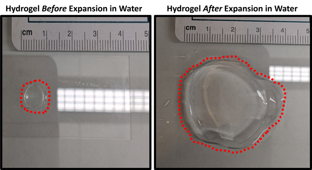
This is where expansion microscopy (ExM) proves beneficial. Rather than depending on optics for magnification, ExM physically increases biological tissues’ size by encasing them in a hydrogel, a water-retaining polymer that can enlarge without distorting its shape — similar to materials utilized in baby diapers. As the hydrogel expands, the cellular structures also grow, enhancing visibility of small details even through a regular microscope. Thus, instead of a close-up image where individual components may appear hazy or warped, the actual size of the cells increases, akin to a sponge soaking up water. Even better, it remains an economical and readily available method.
Although ExM has gained traction in animal studies, its application to plants has posed challenges. The sturdy cell walls composed of cellulose in plant cells obstruct uniform expansion.
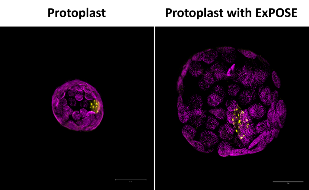
Cox and his team addressed this challenge by utilizing protoplasts — plant cells with their walls eliminated — enabling them to adapt ExM successfully for plant investigations. The outcome is ExPOSE, a technique that offers high-resolution, intricate views of plant cells.
With ExPOSE, researchers can now observe a plant’s cellular structures with enhanced resolution, facilitating exploration of the precise positioning of proteins, RNA, and other biomolecules. This is crucial for Cox, whose research is centered on cellular communication and responses. “This enhances our comprehension of the location of these genes and proteins, their functionality, and their roles in cellular responses,” he remarked.
An innovative new toolkit for plant biology
However, ExPOSE is merely one element of cellular imaging and data acquisition. Cox pondered, “What additional methods could we synergize with this to create more of a toolbox?” When ExPOSE was integrated with techniques such as hybridization chain reaction, commonly referred to as HCR, and immunofluorescence, Cox and his team discovered they could observe both proteins and RNA with even greater clarity.
While ExPOSE is presently utilized for studying individual cells, Cox imagines an even more extensive future for expansion microscopy in plants. “Our aim is to comprehend spatial data at a cellular level and then also, collectively, at a broader scale,” Cox clarified. This signifies utilizing ExPOSE to examine organs, leaves, roots, and ultimately, entire plants, allowing researchers to investigate the intercellular communication.
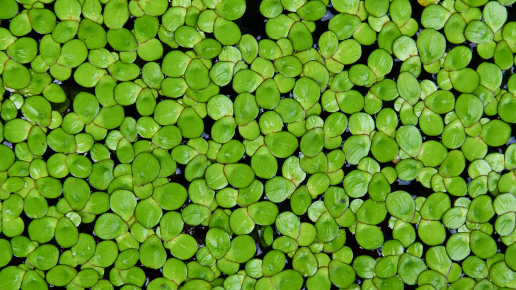
At the core of Cox’s research lies a humble yet potent model organism: duckweed. This petite, fast-growing aquatic plant presents a promising opportunity for probing cellular communication and gene expression. “Due to the compact size of duckweed, it serves as a model to grasp the activities of each cell at any given time,” Cox stated. This proves particularly beneficial when investigating plant cells’ responses to stress, including infections or shifts in the environment.
The overarching aim? To apply this knowledge to agricultural practices. By understanding how plant cells communicate and protect themselves from threats, researchers might cultivate more resilient, higher-yielding, and rapidly growing crops, thus enhancing food security and sustainability.
The post Introducing expansion microscopy to plants was first published on The Source.