Employing a method known as DNA origami, researchers at Caltech have created a technique that may result in more affordable, reusable biomarker sensors for the rapid identification of proteins in bodily fluids, removing the necessity to dispatch samples to laboratory facilities for analysis.
“Our research establishes a proof-of-principle demonstrating a pathway to a single-step procedure that could be utilized to detect and quantify nucleic acids and proteins,” states Paul Rothemund (BS ’94), a visiting associate at Caltech in the fields of computing and mathematical sciences, and computation and neural systems.
A manuscript explaining the study has recently been published in the journal Proceedings of the National Academy of Sciences. The primary authors of the manuscript are former Caltech postdoctoral researcher Byoung-jin Jeon and current graduate student Matteo M. Guareschi, who conducted the research in Rothemund’s laboratory.
In 2006, Rothemund released the initial article on DNA origami, a method that grants straightforward yet remarkable control over the configuration of molecular constructs at the nanoscale utilizing solely DNA.
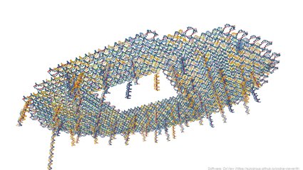
Fundamentally, DNA origami allows lengthy strands of DNA to fold, via self-assembly, into any intended shape. (In the 2006 article, Rothemund famously employed the technique to produce tiny DNA smiley faces measuring 100 nanometers wide and 2 nanometers thick). Researchers start with a lengthy strand of DNA, referred to as the scaffold, in solution. Given that the nucleotide bases constituting DNA bind in a predetermined manner (adenine pairs with thymine, and guanine pairs with cytosine), the scientists can incorporate hundreds of short sequences of corresponding DNA, ensuring they will attach to the scaffold at specified locations. These short, added DNA fragments fold the scaffold and form its structure, acting as “staples” that maintain the integrity of the construct. This technique can subsequently be utilized to produce shapes ranging from a representation of North and South America to nanoscale transistors.
In the latest study, Rothemund and his team employed DNA origami to construct a lilypad-like formation—a flat, round surface roughly 100 nanometers across, connected by a DNA linker to a gold electrode. Both the lilypad and the electrode possess short DNA strands available to bind with an analyte, a molecule of interest within the solution—whether it be a DNA molecule, a protein, or an antibody. Upon binding of the analyte to these short strands, the lilypad gets drawn down to the gold surface, bringing 70 reporter molecules on the lilypad (which signify that the target molecule is present) into proximity with the gold surface. These reporters are redox-active molecules, meaning they can readily lose electrons during a chemical reaction. Hence, when they are sufficiently close to an electrode, an electric current can be detected. A stronger current signifies the presence of a greater quantity of the target molecule.
Previously, a similar strategy for creating biosensors was devised using a single DNA strand instead of a DNA origami structure. That earlier research was spearheaded by Kevin W. Plaxco (PhD ’94) of UC Santa Barbara, who is also a co-author of the current manuscript.
Caltech’s Guareschi emphasizes that the new lilypad origami is considerable in size compared to a solitary DNA strand. “This allows it to accommodate 70 reporters on a single molecule and maintain their distance from the surface until binding occurs. Then, when the analyte is engaged and the lilypad encounters the electrode, a substantial signal amplification occurs, making the change straightforward to observe,” Guareschi explains.
The relatively sizable nature of the lilypad origami also permits the system to readily identify and interact with larger molecules, such as substantial proteins. In the new manuscript, the team demonstrated that the two short DNA strands on the lilypad and the gold surface could function as adapters, transforming it into a sensor for proteins rather than DNA. In their research, the scientists added the vitamin biotin to these short DNA strands to convert the system into a sensor for the protein streptavidin. They then included a DNA aptamer, a strand of DNA that can attach to a specific protein; in this instance, they utilized an aptamer that binds to a protein known as platelet-derived growth factor BB (PDGF-BB), which could assist in diagnosing conditions such as cirrhosis and inflammatory bowel disease.
“We simply add these uncomplicated molecules to the system, and it becomes prepared to detect something different,” Guareschi affirms. “It’s sufficiently large to accommodate whatever you need—whether that be aptamers, nanobodies, fragments of antibodies—and it does not require a complete redesign each time.”
The researchers further demonstrate that the sensor can be reused multiple times, with new adapters incorporated for each different detection. Although the performance slightly diminishes over time, the current system is capable of being reused at least four times.
Looking ahead, the team aspires for the system to also have applications in proteomics—investigations that determine the proteins present in a sample and their respective concentrations. “You could employ multiple sensors concurrently with various analytes, and then you could perform a wash, switch the analytes, and remeasure. And you could repeat this several times,” Guareschi elaborates. “Within just a few hours, you could quantify hundreds of proteins using a single system.”
Additional authors of the manuscript, “Modular DNA origami-based electrochemical detection of DNA and proteins,” include Jaimie M. Stewart of UCLA; Emily Wu and Ashwin Gopinath of MIT, Netzahualcóyotl Arroyo-Currás from Johns Hopkins University School of Medicine, Philippe Dauphin-Ducharme from the Université de Sherbrooke in Canada; and Philip S. Lukeman from St. John’s University in New York.
The team utilized fabrication tools at the Kavli Nanoscience Institute at Caltech. The study received support from the Army Research Office, the Office of Naval Research, the National Science Foundation, and the Life Sciences Research Foundation backed by Merck Research Laboratories.