
“`html
Science & Technology
Stunning revelation
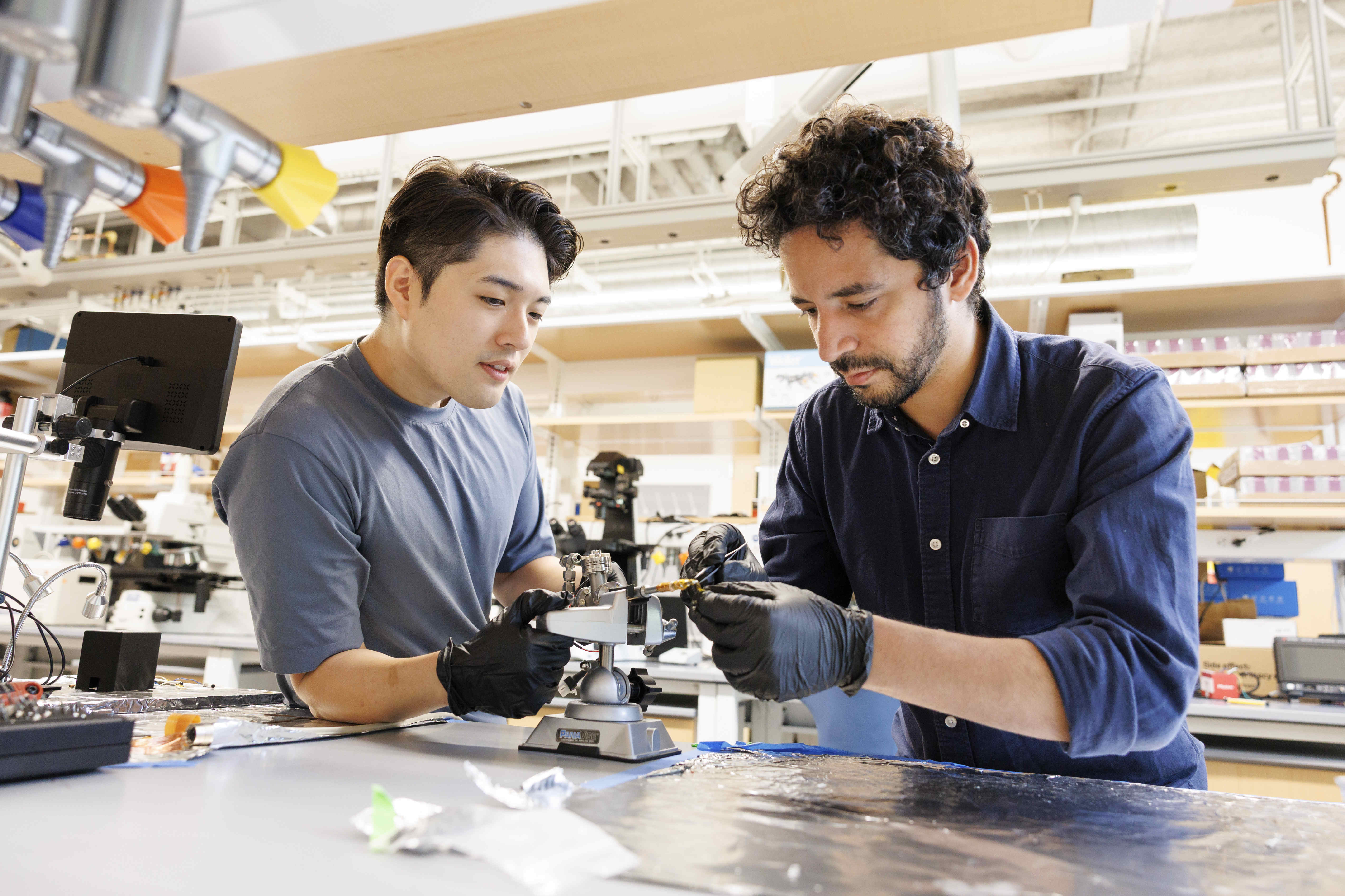
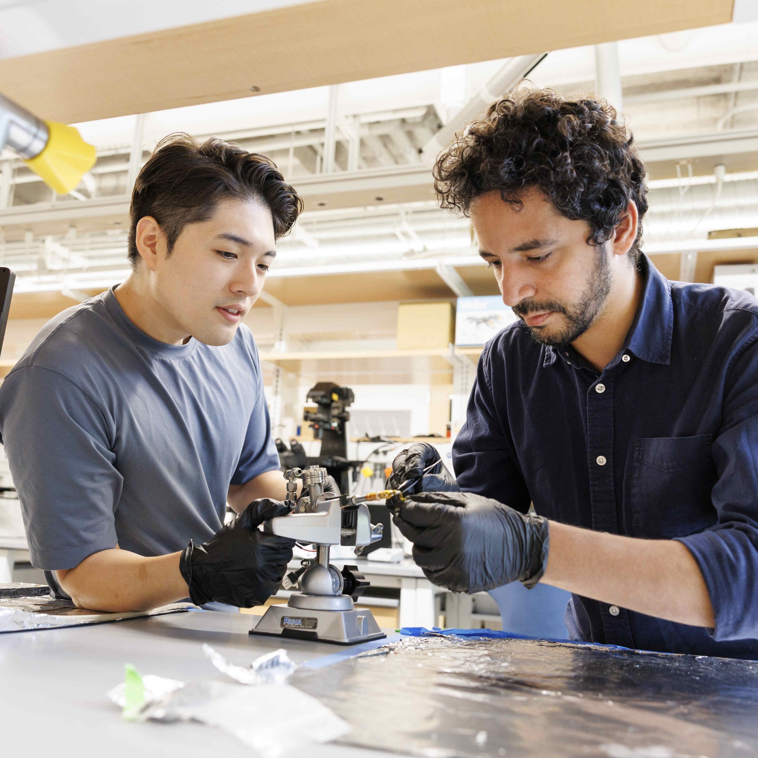
Suk Hyun Sung (left) and Ismail El Baggari.
Images by Niles Singer/Harvard Staff Photographer
Physicists push boundaries to capture quantum substances
Investigators at the Rowland Institute at Harvard have developed an innovative approach to reach the lowest temperatures possible for imaging materials on a sub-atomic level. By merging the specialized expertise of Rowland staff scientists with colleagues at the University of Michigan, these results herald a new phase of ultra-cold microscopy.
Cryogenic transmission electron microscopy — TEM — has historically been crucial in numerous scientific fields, ranging from biology to physics, as the extremely low temperatures enable detailed scrutiny of samples from inorganic crystals to intricate biomolecules at the atomic level. Ordinarily, the cryogen, or coolant, used is liquid nitrogen, which boils at 321 degrees below zero Fahrenheit (or 77 Kelvin) — impressive, but insufficient for observing those odd quantum movements.
Consequently, researchers have aimed over the past ten years to achieve even lower temperatures by employing liquid helium, which boils at 421 degrees below zero Fahrenheit, or 4 Kelvin, bringing it extremely close to “absolute zero.” However, this introduces significant technical challenges that impact the mechanical stability of the microscope.
Harvard scholars conceptualized a novel method to utilize liquid helium for a more secure approach to this advanced microscopy, a significant advancement outlined in a manuscript published last month in PNAS. Spearheaded by principal investigator and Rowland associate Ismail El Baggari, the team formed innovative partnerships to establish a practical new technology, both within the institute and in collaboration with University of Michigan Professor Robert Hovden. This research marks one of the initial high-impact publications (another being robot flies) since Rowland transitioned to Harvard’s science campus a year prior.
The original idea of cryogenic cooling during microscopy to maintain the integrity of biological molecules — which led to the 2017 Nobel Prize in chemistry — was focused on studying cells, proteins, and other soft matter. Nevertheless, El Baggari and Suk Hyun Sung, a postdoctoral researcher in the team, recognized its potential in quantum physics.
“We’re physicists and we are intrigued by imaging unusual materials known as quantum materials that exhibit different properties at low temperatures,” El Baggari expressed, emphasizing that electrons demonstrate quantum behavior only at extremely low temperatures. “Thus, we must cool down the materials to unveil those new attributes.” Utilizing liquid helium cooling was perceived as a solution for this.
For applying liquid helium, however, the researchers required an electron microscope that could enable such cooling. “In prior attempts, we have employed electron microscopy to visualize the atomic structure of these materials at elevated temperatures, yet it turns out that obtaining high-resolution images with liquid helium cooling is quite challenging,” El Baggari remarked.
“Liquid helium evaporates rapidly and effortlessly, as any external heat causes its vaporization,” he noted. “This introduces vibrations as bubbles emerge when the flow is disturbed by a mixture of gas and liquid.”
These vibrations blurred the images similarly to how a bubbling pot of boiling water obscures the view through it, an effect still visible when the water is cold. The rapid evaporation also meant that the low temperatures could only be sustained for about 20 minutes, far too brief for meaningful measurements.
“We developed a geophone for the microscope that is so responsive, it can sense vibrations within fractions of atomic distance.”
Winfield Hill, director of electronic engineering
To address this issue, the researchers needed to devise a technique that would permit the use of liquid helium while preventing vibrations. They collaborated with Rowland manager Erik Madsen; director of electronic engineering Winfield Hill; and Alan Stern, a staff computational scientist. Together, they constructed a system capable of maintaining low temperatures for extended periods. They also monitored and isolated vibrations from the cooling system to prevent them from impacting the specimen.
“We had the broad concepts, yet we were initially unaware of the intricacies of cryogenic design, precision machining, and advanced electronics,” said El Baggari. “We had to create new tools and methodologies to start tackling our challenges.”
“Ismail proposed the use of geophones to detect incredibly minute
“““html
oscillations in the ultra-cold TEM configuration, aimed at recognizing areas to mitigate the ‘interference,’ especially on the liquid helium feed. Therefore, we devised a geophone for the microscope that is incredibly responsive, capable of sensing oscillations within fractions of atomic spacing — precisely the dimension required for this microscope to produce clear images at sub-atomic clarity,” remarked Hill.
The risks were considerable due to the intricacy of the apparatus. “These microscopes represent multimillion-dollar investments, and it was rather daunting to contemplate integrating our own novel instrument into something that had never been trialed,” noted Sung.
Into this vast and fragile apparatus, he elaborated, the constructors needed to design site entry holders that “would be positioned as a glass slide slots into a standard microscope — without causing any damage.” The specimen had to be placed correctly within 25 microns, which is one-third the thickness of a human hair. Any discrepancies meant the specimen could not be visualized, vibrations were excessive, or vacuum conditions were subpar.
“During the initial months of assembly, we were simply measuring commercially obtainable site entry holders, ensuring all the dimensions were accurate,” continued Sung. “We lacked knowledge of what the critical dimensions were since all the holders varied slightly, and we were uncertain about what truly counted.” After three years of evaluating the sample holder and enhancing vibration suppression, the team succeeded in capturing their first high-resolution images in quantum materials while functioning for extended periods at ultracool temperatures.
“Abruptly we were investigating materials that we had historically only been capable of analyzing at ambient temperatures,” stated El Baggari. “We had never been able to image them while they were simultaneously demonstrating the emergent properties that we and our collaborators find intriguing. Now we can effectively utilize the electron microscope as a primary instrument to directly explore these quantum electronic phases in materials.”
Ongoing enhancements of the innovative tool could pave the way for new areas of exploration, such as concurrently applying a voltage to operational devices or manipulating materials through stretching or pressing. “The microscopy studies that we’ve consistently conducted at room temperatures can now be performed at low temperatures where it becomes even more thrilling,” he expressed.
The research highlighted in this article received partial funding from the National Science Foundation and the U.S. Department of Energy.
“`