

“`html
In vitro fertilization has served as the primary approach to tackle infertility for nearly fifty years, yet its live birth success rate remains below 40%. Choosing an embryo with the greatest potential might enhance the success rate, but current methods for capturing time-lapse images of developing embryos have been restricted.
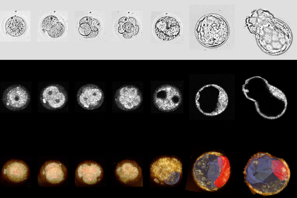
A group of scientists from the McKelvey School of Engineering at Washington University in St. Louis employed dual-modality, 3D, time-lapse optical coherence microscopy (OCM) and brightfield (BF) imaging to observe mouse embryo growth and foresee successful blastocyst formation. Findings from this study, published in Communications Biology this spring, position OCM as a valid technique for selecting high-quality embryos for transfer and increasing the success rates of in vitro fertilization.
This groundbreaking research took place in the laboratory of Chao Zhou, a professor of biomedical engineering and a globally recognized authority in OCM and optical coherence tomography. Contributors to this study include Fei Wang, a doctoral candidate in Zhou’s lab, and Ali Ahmady, an associate professor of obstetrics and gynecology at WashU Medicine.
Zhou is set to present this research at the American Society for Reproductive Medicine’s annual conference in October. Learn more on the McKelvey Engineering website.
The post An inside look at the earliest stage of life appeared initially on The Source.
“`