
“`html
It is now feasible to assess intricate characteristics of electron beams generated by directing electrons to ride on potent laser pulses

In a significant stride toward enabling ultrabright X-ray sources to be more broadly distributed, an international partnership spearheaded by the University of Michigan—with experiments conducted at the U.K.’s Central Laser Facility—has charted essential elements of electron pulses that may ultimately produce laser-like X-ray bursts.
These X-ray bursts offer the potential to propel advancements in chemistry, biology, material science, and physics by allowing researchers to observe molecular behavior in great detail. This method might also find utility in clinical medicine for imaging soft tissues and organs.
Given the incredibly brief duration of these pulses, measured in quadrillionths of a second (femtoseconds), they can capture snapshots of chemical reactions, detailing the choreography of atoms and molecules, including larger biomolecules such as proteins. This research is valuable for both foundational scientific inquiries, extending to quantum mechanics, as well as practical applications in chemistry like drug discovery. The expected influence on the future of science from these compact X-ray free-electron lasers (XFELs) contributed to the funding for this investigation from the U.S. National Science Foundation and Department of Energy, along with various British funding bodies.

“We anticipate that laser-plasma accelerators will be capable of condensing XFELs to the dimensions of a tabletop and substantially enhancing the accessibility of XFEL sources, yet one challenge lies within the quality of the beam. This innovative diagnostic reveals that the beams we generate possess significantly higher quality than earlier perceptions,” remarked Alec Thomas, U-M professor of nuclear engineering and radiological sciences and corresponding author of the study published in Physical Review X. Thomas also holds positions in electrical and computer engineering and physics.
Electrons utilized for producing powerful X-rays are typically generated in accelerators that measure hundreds of meters in length, with just one such facility available in the U.S. and five others dispersed globally, according to Thomas. However, employing intense laser pulses to accelerate electrons may broaden access to the method by utilizing cost-effective, commercially available components and necessitating a smaller lab footprint.
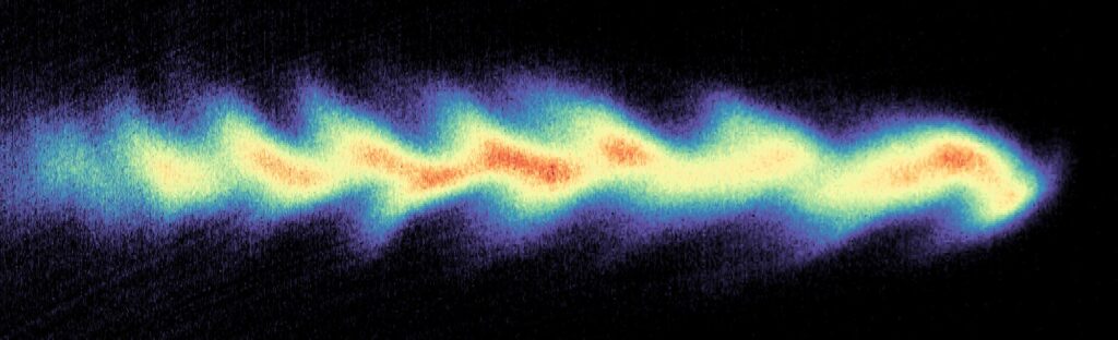
The novel technique involves directing a femtosecond-scale laser pulse through a gas cloud. The light extracts electrons from the gas atoms, and some of these electrons are subsequently drawn along in the laser pulse’s wake, a process termed laser wakefield acceleration. The properties of this electron beam dictate the characteristics of the resultant X-ray pulse. For instance, to yield laser-like X-ray pulses optimal for imaging soft tissues, the electrons must be grouped together in clusters within the pulse.
The international team has illustrated a technique to map the electrons within the pulse, noting their trajectory and velocity. Specifically, they can segment the beam into slices and ascertain the energy distributions within these segments.

“The temporal resolution of our method is roughly one femtosecond, surpassing the diagnostics currently available at state-of-the-art conventional radio-frequency accelerators,” stated Yong Ma, U-M assistant research scientist in nuclear engineering and radiological sciences.
The team discovered how to attain this resolution through an experiment conducted on the Gemini laser in Didcot, U.K. The wave structure of the laser light used for electron acceleration already imprints on the electron beam, establishing a predictable wave pattern. However, the momentum of each electron introduces variations from the anticipated pattern, and the team successfully interpreted those variations to reconstruct the attributes of the electron beam.
The beam was evaluated by redirecting it onto a screen, distinguishing the electrons based on energy and measuring the angle at which each electron impacted. This allowed for the determination of each electron’s momentum while also indicating its original location in the beam. Following this, the team developed a machine learning algorithm capable of utilizing this data to reconstruct the specifics of the original pulse.
This information can be leveraged to refine the characteristics of electron beams in prospective compact X-ray facilities. Continuing their exploration of measuring electron beams produced by laser pulses, the team has another experiment planned at Europe’s Extreme Light Infrastructure Beamlines in Czechia, in partnership with the U.S. NSF. They also aim to apply the new technique on ZEUS, the highest-powered laser in the U.S., located at U-M and financed by the NSF.
The team comprised researchers from the Central Laser Facility, U.K.; Queens University, Belfast, U.K.; Cockcroft Institute, U.K.; Lancaster University, U.K.; Diamond Light Source, U.K.; University College London, U.K.; Imperial College London, U.K.; University of York, U.K.; University of Strathclyde, U.K.; Helmholtz Center Dresden-Rossendorf, Germany; Technical University of Dresden, Germany; Helmholtz Institute Jena, Germany; GSI Helmholtz Centre for Heavy Ion Research, Germany; Institute of Physics of the ASCR, Czechia; Lund University, Sweden; Superior Technical Institute, Portugal; Ergodic LLC, U.S.; Lawrence Livermore National Laboratory, U.S.; and University of California, Los Angeles, U.S.
U.S. funding stemmed from NSF Grant No. 1804463 and Department of Energy Grants No. DE-NA0002372, No. DE-SC0022109, and No. DE-SC0016804. U.K. financial support was provided by the Science and Technology Facilities Council, Engineering and Physical Sciences Research Council, and Royal Society. Additional funding sources included EuPRAXIA, the European Commission’s LASERLAB-EUROPE, EuCARD-2, the Swedish Research Council, the Natural Science and Engineering Research Council of Canada, and the Portuguese Foundation for Science and Technology.
“`