
A lot of us have undergone an X-ray at some point, whether at the dentist’s clinic or the physician’s office. Now, astronauts circling the Earth have demonstrated that it’s feasible to perform an X-ray in outer space. This initiative will assist upcoming space pioneers in identifying and tracking medical issues, ranging from fractures and sprains to indications of bone decalcification, while they are in orbit.
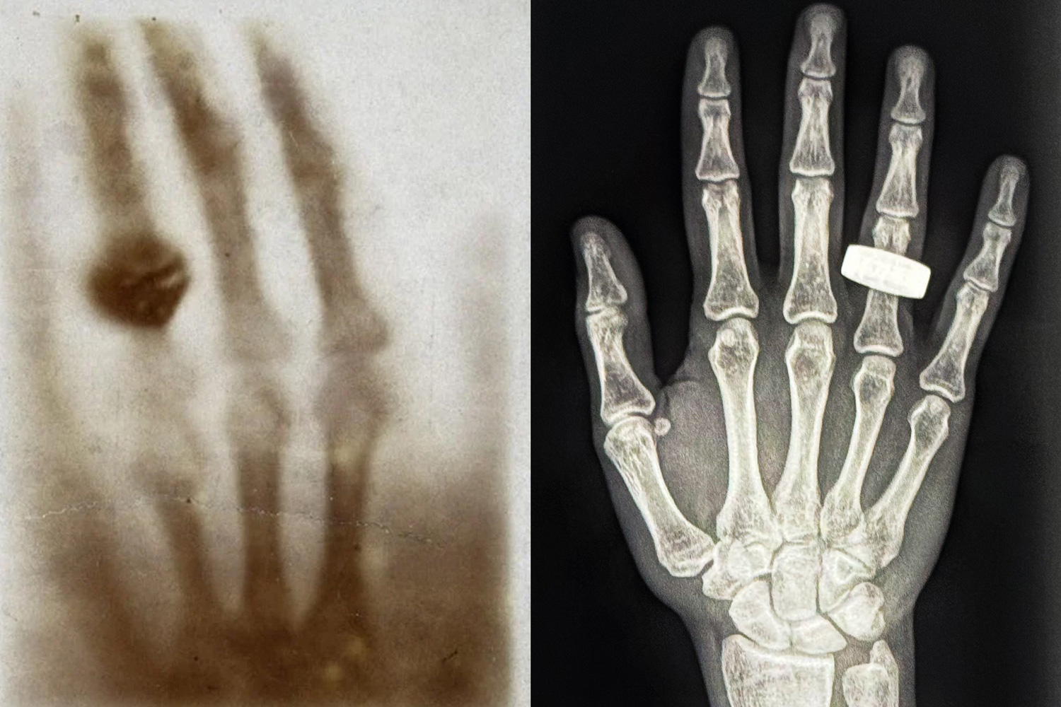
Recently, team members aboard the Fram2 mission took to social media to present the first-ever medical X-ray image captured in space. The photograph is a monochrome scan of a hand adorned with a ring, reminiscent of the initial X-ray image ever created, 130 years ago, by physicist Wilhelm Roentgen, of his wife’s hand. The recent X-ray image was captured in microgravity, inside a four-person spacecraft traveling at orbital speeds of 17,500 miles per hour, approximately 200 miles above the Earth’s surface.
The in-flight body imaging was part of the SpaceXray initiative, which encompassed one of 22 scientific experiments conducted by astronauts during the Fram2 mission. Managed by SpaceX, Fram2 was the inaugural human spaceflight expedition to embark on a polar orbit, circling the planet from pole to pole. The mission name Fram2 is derived from the Norwegian vessel “Fram,” known for being the first to transport explorers to the Arctic and Antarctic regions in the late 19th century.
The body imaging serves as a preliminary demonstration that medical X-ray imaging can be performed within the unique conditions of outer space. Lonnie Petersen, a co-investigator on the SpaceXray initiative, is an associate professor in MIT’s Department of Aeronautics and Astronautics, specializing in space physiology and the consequences of space travel on the human body. Petersen played a key role in defining and developing the protocols surrounding the SpaceXray project, collaborating with institutional partners including Stanford University and the Mayo Clinic, along with X-ray equipment manufacturers KA and MinXray. Petersen spoke with MIT News about how these pioneering in-orbit X-ray images could facilitate the successful and healthy continuation of long-term missions in space.
Q: What difficulties arise when taking an X-ray in space compared to on Earth?
A: There are numerous challenges involving the equipment, techniques, and subjects being imaged.
To have equipment approved for spaceflight, it must be compact and as lightweight as possible. Additionally, there are heightened safety protocols since all devices operate within a confined area. These elevated requirements propel technological advancement. I often claim that space serves as our best catalyst for technology — and this holds true for medical technology as well.
For this endeavor, we utilized a portable, specialized X-ray generator and detector designed by MinXray and KA Imaging for battlefield applications, adapting it for use in space.
Regarding methods, I was initially concerned that heightened background radiation might degrade the image quality to a level below clinical standards. However, the first images we have received from space indicate that the quality is excellent. I am eager to conduct a more in-depth analysis of the entire set of images.
We aim for the X-rays to pass directly through the body part in question. This necessitates precise alignment of both the equipment and the subject. As one might expect, positioning a floating subject proves to be more challenging. We will quantify any potential effects of this and incorporate that information for future improvements.
Furthermore, we do not have radiologists or technicians in space. The procedures must be straightforward and resilient enough for a non-specialist to handle.
Finally, regarding subjects: Travel to space significantly affects the human body. Blood and fluids are no longer drawn downward by gravity; instead, they become evenly distributed, leading to changes in local pressures and perfusion. The cardiovascular system and brain experience changes over time. The mechanical unloading of the body results in muscle atrophy and bone decalcification, along with reduced exercise capacity. Given that this mission lasted only 3.5 days, the crew probably didn’t experience many negative effects. However, with an X-ray, we can now assess bone health in space, something we’ve never been able to do before. We can also evaluate potential fluid accumulation in the lungs or investigate illnesses in the abdomen.
I’ll also switch from my physician role to my engineering perspective: X-rays serve as a valuable tool for nondestructive testing of hardware in aviation (and other fields). This project enhances our diagnostic capabilities in space, not only for human subjects but also for equipment.
Q: How did the Fram2 crew manage it?
A: The crew was trained to take X-rays within a single afternoon. It was executed using a train-the-trainer approach. The protocol was established beforehand, and the crew recorded images of one another, verified the quality, and stored the resulting images. We have seen only one image up to this point, but we are truly impressed with the quality, skills, and commitment to advancing science demonstrated by the crew.
Q: What insights will you gain from these initial images?
A: First and foremost, this was a technology demonstration: Is it even feasible to perform this in space? We are eager to analyze all the images, but preliminary data indicates that we absolutely can. Next, we will conduct a detailed evaluation to extract all the insights possible concerning current capabilities as well as future steps. The team is certainly enthusiastic about pushing this research forward and exploring even more groundbreaking developments.